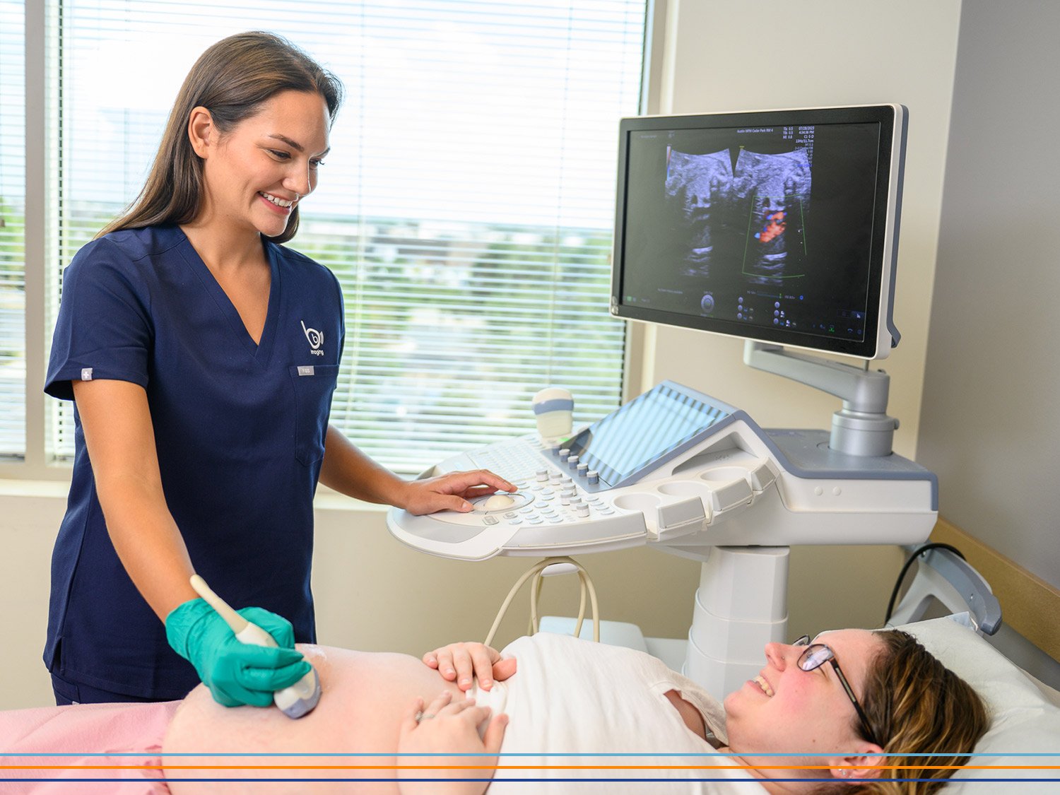
This one is for sonographers. We want all of you to take care of yourselves and enjoy long, fulfilling careers. Pain and injury creep into this industry so quickly, and we know many of you have experienced or are currently struggling with work-related musculoskeletal disorders (WRMSDs).

So, in honor of the upcoming holiday, we’re handing out three tricks and one lovely treat to help you scan pain-free!

Impact of Sonographer Injuries

Research shows sonographers can experience the onset of WRMSD symptoms as early as six months into their first job. A 2009 study on WRMSDs among U.S. sonographers discovered up to 90% of you are scanning in pain, with all 90% reporting shoulder pain, 69% reporting low back pain, and 54% reporting hand and wrist pain. A 2017 study on WRMSD impact among Chinese sonographers revealed even worse outcomes: 99% of sonographers had experienced WRMSD symptoms. Among respondents, 84% reported shoulder pain, 82% reported lower back pain, and 81% reported wrist and hand pain.

This pain isn’t just impacting sonographers, though. A Society of Diagnostic Medical Sonography whitepaper shows that WRMSDs cost employers more than $120 billion every year in direct and indirect costs. Patients are also impacted as 20% of sonographers suffer career-ending injuries, which takes specialized skills out of the field and contributes to the sonographer shortage.

All that said, we don’t just want you to take care of yourself—we need you to.

Trick #1: Practice Healthy Ergonomics

Ergonomics refers to how people function within their workplace. Your environment has a big impact on your work performance, so it’s best practice to modify the spaces where you work to promote a long and healthy career. As a sonographer, you can set yourself up for success at the beginning of every exam. Don’t rush through or skip this step—this is vital prep work!

Start by adjusting the height of the patient’s bed or chair and asking them to move closer if needed. Next, move the console so that it is close to you, and you aren’t twisting your body to reach it. Now adjust the screen height. It should be at eye level and shouldn’t require you to twist your neck to see it. If your patient doesn’t have a screen to watch and wants to see what’s going on, tell them you need full use of the screen during the exam and assure them they will see their images later.

We know your focus will be largely taken up during the scan but try to check in with yourself occasionally. Do you keep having to fight cords? Consider investing in a cable brace to keep them out of the way. Are you straining your eyes? Try shifting your focus from the screen to objects that are farther away throughout the scan. You might also consider getting a pair of glasses that filter blue light.

Between scans, take as much time as you can to stretch or walk around. Even a few minutes throughout the day will make a big difference.

Trick #2: Adjust for Good Posture and Positioning

Now that your environment is as comfortable as you can make it, it’s up to you to make good choices in how you use it. This is especially true for sonographers when it comes to maintaining proper posture throughout exams. Here are our recommendations:

Hand/Wrist

  • Use a palmar grip rather than a pencil grip.

  • Use a light grip. Your knuckles and fingertips should never turn white from pressure.

  • Keep your hand and wrist in a neutral position and avoid extreme flexion of the wrist.

  • Try alternating which hand you scan with to balance the load.

Shoulders

  • Keep both arms as close to your body as possible.

  • Relax your shoulders down rather than hunching them up toward your ears.

  • As much as possible, avoid raising your arms in any direction. Ergonomics will help with this!

Neck

  • Keep your neck in a neutral position.

  • Adjust the monitor to sit at eye level to avoid hunching over or craning your neck.

  • Keep the monitor facing you to avoid twisting your neck.

Back

  • Try completing some scans standing up. Doing so can relieve pressure on your spine.

  • If you do stand, be sure to adjust the workstation and bed height and consider adding an anti-fatigue mat under your feet.

  • Sit or stand straight up.

The more you keep these tips in mind, the more you can build healthy posture habits that you won’t even have to think about. However, we know it isn’t always possible to practice the best posture and positioning, and that you are very busy. So, in the likely event that your posture suffers, give yourself some grace and go get a massage. You can also avoid pain from bad posture by working out and strengthening those areas of the body and making sure to take time to rest and recover.

Trick #3: Consider a Telesonography® Position

Telesonography provides career optionality and gives you the chance to help more patients. We think it’s going to be a game-changer for WRMSDs in our industry. At BB Imaging, an OB Telesonographer® provides virtual diagnostic services to our physician partners via our telemedicine solution, TeleScan®. In this role, you work from home, which gives your body a break while keeping your specialized knowledge in the field. Not convinced? Check out even more ways telesonography benefits sonographers.

If you find this opportunity as intriguing and exciting as we do, we hope you’ll apply. We’ll be hiring both full-time and part-time roles in 2024, with PRN roles coming soon.

Treat: Take Time to Stretch!

We promised you a treat, and here it is! If you’re eyeing a long career, stretching is key to keeping your muscles limber, avoiding pain and injury, and staying healthy well into the future. That’s why we’re sharing our Dynamic Stretches Poster as a free download. This was created especially for sonographers and includes eight different stretches you can easily do in-clinic or from the comfort of home.

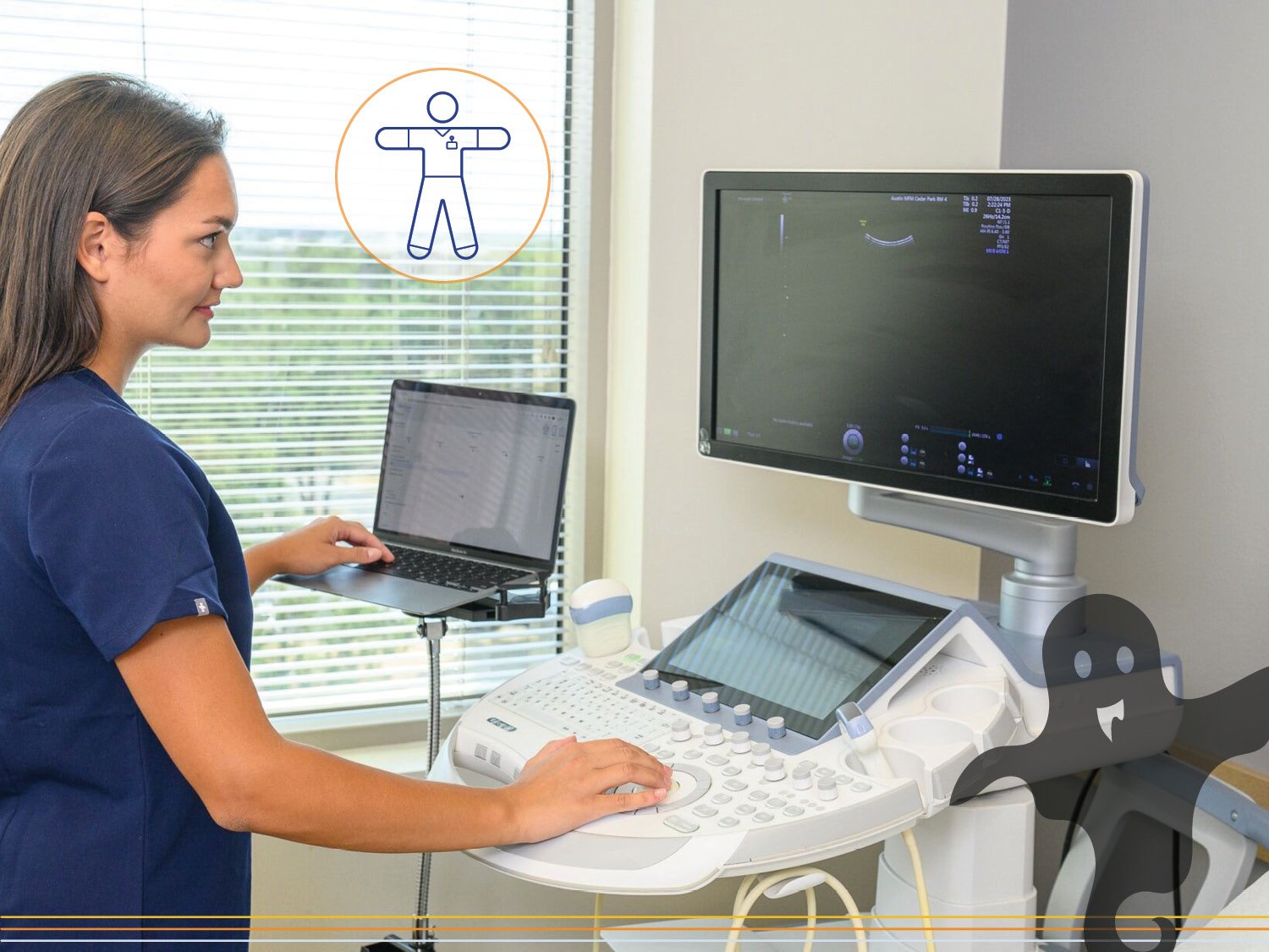
Dynamic Stretches Poster Download

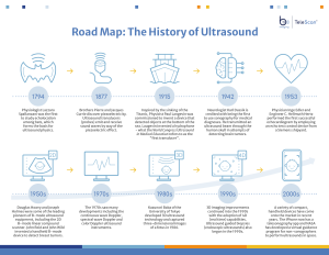
Happy Ultrasound Awareness Month! This month is dedicated to recognizing and appreciating ultrasound technology and the sonographers who wield it. In honor of the occasion, we’ve pulled together a timeline of the history of this increasingly vital imaging modality and explored the depths of innovation in this exciting field.

Feel free to share this timeline to spread awareness of the industry and showcase your place in its storied history.

1700s

Yes, ultrasound is more than 300 years old! In the late 1700s we discovered bats rely on their ears rather than their eyes for navigation. This was an early acknowledgement of echolocation: the location of objects by reflected sound. Similar to bats, ultrasound technology uses sound waves to generate images from the deflected echoes of inaudible, high-frequency sound waves.

1800s

In the late 1800s, the Curie brothers discovered the capacity of crystals to both generate and receive pressure waves in the range of megahertz frequencies, which paved the way for modern-day transducer technology.

Early 1900s

Following the sinking of the Titanic, a Canadian electrical engineer named Reginald Fessenden created an ultrasound-based collision avoidance system as well as sonar-based submarine navigation.

1940s

Ultrasound transitioned to medical applications during WWII. Dr. Karl Theodore Dussik published the first work on medical ultrasonics in Austria in 1942 after using ultrasound to investigate the brain. This experiment was one of the earliest attempts to depict an organ in vivo (Latin for “within the living”).

Dussik used a process known as through-transmission ultrasound, in which a probe on one side of the patient transmits an ultrasonic pulse to a receptor probe on the other side. Due to its limitations, the through-transmission technique would soon be replaced.

Early 1950s

Researchers across the USA, Japan, and Europe developed pulse-echo ultrasound technology. In pulse-echo ultrasound, a transducer both produces the transmitted sound wave and receives its reflected echo. The pulse-echo method is used in multiple ultrasound imaging modes, including:

  • A-mode: A-mode ultrasound imaging is the simplest type of ultrasound and returns echoes in a one-dimensional, graphical format. Usually, it was conducted by placing transducers on both sides of a patient who was partially submerged in water. This method is complex and can’t determine direction or the shape of an object, which has made it nearly obsolete today.

  • B-mode: B-mode ultrasound imaging returns greyscale two-dimensional images. This method added directionality to A-mode data. As it improved, the ability to create multiple B-mode images in rapid succession allowed for real-time imaging and the recording of cine clips.

  • M-mode: M-mode ultrasound imaging displays one-dimensional images over time and is commonly used in cardiac and fetal cardiac imaging to evaluate heart motion.

1955

A doctor used industrial ultrasound technology at a boiler fabrication plant “…to test whether it could differentiate between tissue samples (including an ovarian cyst and a juicy steak).” Spoiler alert: It could. Furthermore, he discovered that when ultrasound was applied to a pregnant person’s abdomen, the technology produced “…a dark oval with crackling shadows.” This imaging offered a window into the uterus, with white lines indicating a placenta in formation, and captured a fetal heartbeat.

1958

A medical article titled “Investigation of Abdominal Masses by Pulsed Ultrasound” was published by Ian Donald, John MacVicar, and Tom Brown. For the first time, ultrasound echo was used for the dating of a pregnancy, which was achieved by comparing current fetal size with fetal growth trajectory charts.

Initially, ultrasound technology within the prenatal space was met with controversy and opposition, as many felt it prioritized scientific rationalization and machinery over centuries of maternal knowledge and intuition.

1963

Midwives and pregnant patients experienced the first obstetric ultrasounds performed by Donald and his colleagues in Glasgow hospitals between 1963 and 1968. The experience was positive, leading to the expression of wonder and delight by expectant parents.

Donald, MacVicar, and Brown produced the “Diasonograph,” the world’s first commercial ultrasound scanner.

1970s

The development of the microchip led to even more technological advances in imaging capabilities. Meanwhile, the price of machines began to drop, which brought ultrasound imaging to the masses.

Doppler imaging became mainstream with the development of color Doppler imaging, spectral Doppler imaging, and continuous-wave Doppler imaging. These methods showcase the movement of blood within blood vessels.

1980s

Kazunori Baba at the University of Tokyo developed 3-D scanning, and patients began to bring their partners and family members to ultrasound appointments, viewing them as exciting life events. To this day, patients receive ultrasound images following their appointments, which is said to contribute to maternal-fetal bonding.

1990s

Power Doppler imaging is added to the mix, which allows for even more detailed studies.

Today—The Future

As you can see, ultrasound has come a long way since the 1700s, and it’s a field that continues to experience innovation. We believe telesonography® is the next big step in the ultrasound timeline—and that it’s happening right now! See how this newest development will impact key demographics by clicking the links below:

And finally, thank you to everyone who participates in this incredible field of medical care. Happy MUAM!

HistoryofUltraSoundFullTimeline_101923

Full timeline can be downloaded here.

Telesonography provides ultrasound services via a telecommunications link. It applies the telemedicine approach to sonography, and it comes with a host of benefits.

BB Imaging is the proud owner of TeleScan®, our very own telesonography solution that connects healthcare providers with expert remote sonographers. We’ll refer to TeleScan and some of the results we’ve seen so far throughout the article. If you’d like to learn even more about it, you can visit our website.

Our mission with TeleScan is to bring prenatal ultrasound care to places where it isn’t already available and accessible—and improve outcomes for pregnant patients across the nation. Those are the driving forces behind our innovation, but we also know this technology can serve as a revenue builder for our provider partners.

Here are seven ways telesonography can impact a provider’s bottom line:

1. Increased Billing

Our partners have realized an average 30-40% increase in billing. This is because medically indicated ultrasound exams can be billed at a higher frequency when they are reviewed by highly credentialed perinatal sonographers. Our telesonographers all hold active registries in OB/GYN and Fetal Echocardiography through the ARDMS and are credentialed in Nuchal Translucency, Nasal Bone, and Cervical Length Education and Review (CLEAR). They all also have a minimum of two years of perinatal experience.

These experts work in TeleScan to provide reports that are both highly accurate and thorough. Data from TeleScan has shown:

  • Exams visualize critical anatomy with greater than 90% reliability in all three trimesters.

  • Remotely estimated fetal weights are well within the established 20% margin of error.

  • TeleScan can identify high-risk indicators and demonstrate the need for further testing.

  • No repeat exams have been required.

At this level of exam quality, our clients have seen that utilizing one room twice a week for TeleScan exams yields an estimated $19,000+ in monthly revenue.

2. Exam and Reporting Efficiency

TeleScan software and workflows are designed to increase clinical efficiency. It takes a healthcare worker an average of 30 minutes or less to capture all necessary cine clips for a TeleScan exam. After capture is complete, a telesonographer® can provide a preliminary diagnostic report in one hour or less—for one facility, this amounted to a 61% decrease in report turnaround times.

What do our partners do with all those time savings? They schedule additional patient appointments. On average, the increase in available time slots cuts the time until the next available appointment by 50-75% within just a few months of service.

3. Reduced Recruitment Costs

As an ultrasound service provider, we’re keenly aware of how difficult it is to hire sonographers. A longstanding shortage makes an already small talent pool even smaller, and issues like injury and burnout can take sonographers out of the field early in their careers. Currently, the recruiting cycle can take 180+ days to hire for an open role. In addition, many facilities are now offering sign-on bonuses to compete for limited talent, making the hiring of a sonographer an increasingly expensive endeavor.

Through a TeleScan implementation, you can access perinatal sonographers in as little as one month—no hiring required.

4. Reduced Retention Costs

As we mentioned previously, pain and injury play a big role in sonographers quitting the field. If you already have onsite sonographers, we’re sure you know long days of scanning can bring on work-related musculoskeletal disorders, which can result in high turnover rates and career-ending injuries.

Telesonography is designed to take some of the physical burden of scanning from these hard-to-find professionals. TeleScan can augment care for low-risk pregnancies while allowing your onsite sonography experts to focus on more complex or high-risk examinations. This offers your sonographers a balanced schedule and the capacity to work at the top of their license, which can lead to more years of happy, healthy scanning.

5. Continuity of Care

If your facility doesn’t currently schedule ultrasounds in-house, we’re guessing your patients have to travel to third-party imaging centers. From scheduling (and rescheduling when the patient no-shows) to chasing down reports, we know this setup can cause a lot of hassle for both you and your patients.

Telesonography provides the option to keep patients in-house for their imaging, which increases patient compliance, and allows you to offer a seamless patient experience. Minimizing frustration can also lead to higher patient satisfaction and retention.

6. Appropriate Escalation of Care

When you keep patients in-house for imaging, you can also provide immediate escalation of care when necessary. If TeleScan discovers pathology that requires immediate attention, we will follow a pre-determined plan to inform the right people, through the right channels. This way, the patient can receive the care they need directly from you.

7. Expanded Reach

Telesonography is an excellent solution for expanding your reach, especially if you can take advantage of underutilized outreach clinics and satellite locations. This is especially helpful for patients who live in remote areas or face transportation challenges—and can be advantageous to many more.

For example, one of our partners utilized TeleScan in a satellite clinic and provided a shorter travel time for 20% of patients who were currently driving to the main campus. Another partner decreased their patients’ average distance to ultrasound from 120 miles to just 5 miles.

That all sounds good… but how does it work?

TeleScan is a software-as-a-service product that connects healthcare providers with expert remote sonographers.

Here’s how it works: A knowledgeable healthcare worker (like a medical assistant) uses TeleScan and an ultrasound machine to capture cine clips, which are uploaded to our secure cloud platform. From there, a remote, perinatal sonographer reviews them, provides analysis and annotations, and compiles a preliminary diagnostic report. When the report is ready, the provider is notified, and can use TeleScan to review, update, and finalize the report. If they have opted in, the patient will receive selected keepsake images via a secure text message.

TeleScan is designed to be accessible and financially feasible for everyone. It utilizes existing resources, works with 2D machines, and is paid via a tiered monthly subscription.

Ready to learn more?

Watch Telescan® Demo

Austin, Texas — April 4, 2025 — BB Imaging has been named to the 2025 Inc. Regionals Southwest list, ranking No. 52 among the fastest-growing privately held companies in the region. This marks a significant jump from its previous ranking at No. 131 in 2023, reflecting a 126% two-year growth rate.

This prestigious recognition, published by Inc. magazine, honors companies that have demonstrated exceptional expansion and impact. This year’s Southwest honorees contributed more than 13,800 jobs to the U.S. economy and generated a collective $15.9 billion in revenue, with a median growth rate of 106% from 2021 to 2023.

Founded in 2005, BB Imaging is celebrating 20 years of advancing equitable access to high-quality ultrasound care. The company provides diagnostic imaging solutions to underserved communities and healthcare systems nationwide through scalable staffing, clinical expertise, and patient advocacy.

“This recognition by Inc. highlights our commitment to delivering compassionate, expert care in places that need it most,” said Blanca Lesmes, President, Chairperson, and Co-founder of BB Imaging. “For 20 years, we’ve been pushing boundaries—geographic and systemic—to ensure every person, no matter where they live, has access to the care they deserve.”

“This milestone isn’t just about growth, it’s about impact,” added Sanjay Patel, CEO of BB Imaging. “Every scan, every patient, every partnership is part of a larger mission to change healthcare for the better.”

BB Imaging continues to champion innovation and accessibility in healthcare by investing in their sonographers’ professional development, elevating the industry, and transforming care delivery models to close gaps in coverage.

The complete list of Inc. Regionals: Southwest winners is available at inc.com/regionals/southwest.

About BB Imaging: BB Imaging delivers high-quality sonography services designed to improve health outcomes and expand access to care. We partner with healthcare facilities to provide expert diagnostic ultrasound with a compassionate touch, carried out by skilled sonographers who see their work as both science and service. Our scalable model eases operational burdens, helping partners focus on patient experience, because we believe everyone deserves exceptional imaging and the dignity that comes with it.

Media Contact: Jennifer Whisnant / Email: jennifer.whisnant@salesfactory.com

To meet our mission

As a team, we’re working to improve health outcomes by making high-quality ultrasound services accessible to everyone. This mission has defined us since Day One, and it tops our wish list because we remain totally committed to it. Right now, we’re pursuing this goal in two ways: onsite ultrasound services and telesonography®.

We started with our onsite services 18 years ago, and they’re still going strong. Our dependable and highly credentialed sonographers work with provider partners across the United States to provide access to a variety of ultrasound modalities. Our partners have found us to be helpful, reliable, and trustworthy, which has given us a great reputation in the industry.

Just this year, we launched our new telesonography service, TeleScan®. It’s a secure software that connects providers and their patients with remote sonographers. This solution came about because we kept seeing the need to reach patients who can’t access bedside ultrasound care. More than 50 of our sonographers helped to develop this innovative product, and now it’s seen more than 1,000 patients.

The best thing about this wish list item is that we’re not just wishing for it… we’re working toward it every day!

To keep learning

Our team is dedicated to excellence. We desire to be the best at what we do, so continuing to add to our knowledge and accomplishments is a wish that is near and dear to our hearts.

We’re not seeking accomplishment for only ourselves though. We’re also hoping to elevate others and gladly share what we know with you through our webinars, newsletter, and blog posts.

We hope you’ll take advantage of these free resources and let us know if there’s another topic you’d like to hear about.

To make more friends

Our team members tend to form close-knit communities—and we love it that way! That’s why our third wish is to keep that going by adding folks to our team. All across the country, we need more people to help us meet the demand for ultrasound services.

By the way… we’re not a staffing agency (but we get that a lot). You will be on OUR team and that comes with some sweet benefits that are specifically designed to meet the needs of clinical workers:

  • Monthly wellness benefit: We provide a monthly stipend for you to spend on whatever makes you feel well—massages, gym memberships, yoga classes, etc.

  • PTO on your birthday: We all need to feel loved and celebrated, and our gift to you is a full day of PTO to use on or around your birthday.

  • The schedule you want: Wanna travel? We have roles for that. Looking for regular work hours? We have roles that are never on-call and get weekends off.

If you like those benefits, you can see the rest by viewing our job listings.

If you’re not interested in a new job, we hope you’ll be our friend on social media. Follow us on Facebook, Instagram, and LinkedIn to get a peek into our culture, stay updated, and participate in giveaways.

To add something new to the trophy case

Our final wish is to earn a shiny new acknowledgement next year.

This year we were honored as one of the fastest-growing companies, both in Inc.’s Southwest Region and nationally on the Inc. 5000 list. We also won a Silver Stevie in the American Business Awards and were honored with a writeup on their website.

Our Cofounder and CEO, Blanca Lesmes, also bagged a couple of high-profile awards, including Inc. Female Founders and a Woman’s Way Business Award.

And just last week, TeleScan won the Technology Innovation category in the Fierce Healthcare Innovation Awards, which identify and showcase innovation that is improving and transforming the industry.

These are the things on our Christmas list, but what about you? What do you hope the holidays and new year will bring for you?

In the clinic and beyond, sonographers have many opportunities to use their skills, expand their horizons, and grow their careers. In this article, we’ll look at clinical options that range from charge sonographer all the way up to vice president of clinical services. Along the way, we’ll suggest steps you can take to grow into each role successfully.

Clinical Jobs for Sonographers

If patient care is your passion, it’s entirely possible to stay on the clinical side of the industry while growing your career. Consider this hypothetical career trajectory: from sonographer to charge sonographer, to department supervisor, to practice manager, to vice president of clinical services. The titles may be different in the health system where you work, but similar roles are likely available.

And if the path we suggest seems impossible, you may need to work with a company that better supports your growth. BB Imaging is a big believer in internal promotions. Join us and start building your dream career today.


How to become a charge sonographer

Charge sonographers scan patients and take on additional leadership responsibilities like quality assurance tasks, continuing education, and training new team members.

If that sounds like something you’re interested in, consider taking these steps to set yourself up for success:

  1. Gain scanning experience and regularly ask for feedback: Make it your goal to have a reputation for being “that sonographer”—the one who pursues ultrasound excellence with the passion of a Taylor Swift fangirl. Ask to do the scans you know you need practice in and keep learning. One of the easiest ways to learn is to ask for feedback 🔁.
  2. Add a registry or credential: One of the easiest ways to set yourself up for leadership is to become multi-certified. Check out the ARDMS certification list to see what other areas you’re interested in, and then stop by our tips and tricks blog 🤓 for advice on acing your next board exam.
  3. Mentor younger sonographers: As you gain experience, remember to elevate the next generation of sonographers. Your expertise is invaluable, and even one year in-clinic can set you up to be a great mentor 👩‍🏫 for a fresh graduate.
  4. Learn to communicate effectively: Communicating with patients, team members, supervisors, and physicians is a big part of a sonographer’s job. Learning to communicate effectively can include becoming more compassionate in your speech, onboarding physician preferences, or even advocating for yourself 💪.


How to become an ultrasound department supervisor

In this next step up, you may add accreditation and schedule management tasks and even be responsible for helping to meet financial goals.

So, while you’re in your charge sonographer era, here are some career-building steps to prepare you for a supervisor role:

  1. Stay current on best practices and new technologies: Ultrasound is developing and changing rapidly, so staying updated is essential. Sign up for an AIUM membership and industry newsletters (like ours 👀) for delivered-to-you updates. And don’t neglect industry conferences or online events like our webinars that help you stay sharp.
  2. Build rapport: It’s a no-brainer to promote someone who gets along with everyone else. You can be that person! Here are eight ways to start building rapport with your team.
  3. Demonstrate your organization: Organizational skills take many forms. Are you a great time manager? Can you recognize others’ skill sets and delegating tasks effectively? Then speak up, take on those tasks, and showcase your talent!
  4. Push past the status quo: “The way we’ve always done it” might not be the best way to do it anymore. Someone willing to ask questions and suggest improvements demonstrates their readiness for greater responsibility.


How to become a practice manager

Practice managers are still heavily involved in clinic life but take on more administrative tasks. This role is a great growth opportunity for sonographers looking to remain in healthcare without aggravating a musculoskeletal injury. As a practice manager, your workload may include managing clinic processes, hiring and retaining team members, and managing budgets.

Here are some ways to stand out as a great fit for this role:

  1. Own departmental projects: As a department supervisor, you’ll likely gain experience creating and maintaining processes. Use that time to prove that you take initiative, have leadership skills, and can communicate effectively to accomplish goals.
  2. Gain interview experience: As time allows, sit in on new hire interviews or prospective employee assessments. Get familiar with the signs of high performers and collaborative team players.
  3. Foster cross-departmental relationships: Working in cross-departmental teams shows you understand how all the pieces work together to create an excellent patient experience.
  4. Earn an advanced degree in healthcare administration: As you move into more administrative-heavy work, it can be helpful to pick up new knowledge and terminology and add new perspectives to your toolbox.


How to become a vice president of clinical services

In this role, you will develop, manage, and report on new programs, choose and implement enterprise-wide technical systems, and interface with C-suite leaders.

Here are some ways to set yourself up for success in this role:

  1. Tell your success stories: Use the skills and experience you’ve gained from previous roles to position yourself as a qualified candidate. If you’ve done even a few of the tasks we’ve recommended already, you have a great list of accomplishments to add to your resume.
  2. Master communication skills: As a VP, you’ll be responsible for organizing teams, getting people on the same page, and overseeing change management—all tasks that require effective interpersonal skills. Pro tip: start practicing these now (in whatever role you hold), and they’ll be second nature when you reach this level!
  3. Build and leverage relationships: They say, “your network is your net worth,” and they’re not wrong. Industry organizations, product and service vendors, and healthcare educators can lend a lot of support to the goals you’ll be chasing, so start building those relationships now.
  4. Fill in the gaps: We’ve presented a hypothetical career path, but titles and responsibilities will vary. If you’ve missed anything along the way, find a course or class (or, better yet, find a mentor!) to build your knowledge in that area.


Get started—become a sonographer!


Welcome to part two of our deep dive into career paths for sonographers! If you missed part one, we discussed the clinical roles sonographers can grow into. In this article, we’ll take a look at other ultrasound industry jobs as well as potential roles in sales and product development. Some of these options may surprise you!

Ultrasound Industry Jobs

If you’re ready to say “Bye, Felicia” to the clinic without exiting the ultrasound industry, we’re happy to share that there are plenty of options for you to explore!


How to become an ultrasound educator

Ultrasound educators are vital to the industry. With an ongoing shortage in this highly specialized field, we need high-quality educators more than ever.

Here’s how to start adding educator skills to your skillset:

  1. Get comfortable speaking: Public speaking is a large part of a teacher’s role, so it’s a good idea to gain as much confidence as possible before making a long-term commitment to a school.

  2. Get organized: Other educator tasks include building curriculums, managing class schedules, and posting grades. If you can read between the lines, you’ll see that those tasks mean you’ll need to do a lot of organizing—of your time, tasks, and resources.

  3. Interpersonal communication skills: The teachers who make the most impact on students are the ones who communicate best. Whether that means making lessons more engaging or connecting on a personal level, improving your classroom communication will make you a standout contributor to your students’ careers.

  4. Consider an advanced degree in sonography: Earning an advanced degree will help you stand out among applicants and smooth the road to promotion within the educational sector.


How to become a DMS program director

Speaking of which… how would you feel about becoming a diagnostic medical sonography program director? This person oversees the creation and maintenance of the program, hires faculty, and forges strong industry connections.

Here are some ways to get a head start:

  1. Start with adjunct or faculty roles: Experience as an educator will help you better understand how the program works. You’ll also be able to identify opportunities for improvement, which you can take with you into a director role.

  2. Stay updated: Accreditation paperwork isn’t the most dazzling part of the role but is vitally important. You’ll also need to keep an eye on regulatory changes. Look at it this way – you’ll always be on the leading edge of ultrasound practice changes!

  3. Build relationships: Industry connections will be vital in this role, so we hope you’re a people person. Among other things, you’ll partner with healthcare facilities and sonography companies to provide clinical hours for students and job opportunities for new grads.

  4. Build your network: This is so important that we listed it twice. Consistently making new contacts will be vital to the growth of your program, so get ready to shake hands with ultrasound industry organizations, community groups, recruiters, and more.

🤝 By the way… did you know we work with DMS directors from all over the country? It’s true! Contact us to learn more about how we can help each other.


How to become an ultrasound researcher

You don’t have to pursue this avenue exclusively—you can become a published researcher in whatever role you hold!

Let’s break down some steps to take that will make this path easier:

  1. Become a reviewer: Publications are always looking for reviewers. Reviewing others’ work will familiarize you with the language, format, and expectations for authors.

  2. Start where you are: Did you know new research isn’t necessarily required to become a published author? It’s true! Try a literature review. Your job will be to pull new insights or make a new argument based on previously published information.

  3. Gather requirements and submit: Before you start your study, review the requirements for the journal you’re targeting and the type of article you plan to submit. If you’re looking for potential journals to submit to, here are a few: Journal of Diagnostic Medical Sonography, American Journal of Obstetrics and Gynecology, Cardiovascular Ultrasound, and Journal of Clinical Ultrasound.

  4. Onboard feedback: Reviewers can seem picky, but they’ll help you achieve the highest standard. You’ll know about this firsthand if you start as a reviewer yourself! Remember not to take it personally. Just make the suggested edits and resubmit.

How to become a public speaker

Let us tell you a secret: all it takes to be a great public speaker is practice! Anyone can learn to share their knowledge from a stage, and as an expert sonographer, we know you have something worthwhile to share.

Here’s how to become a dynamite speaker:

  1. Get comfortable on a stage: Most people fear public speaking, but it gets a lot easier when you start small. Get some practice by joining a program like Toastmasters or presenting to coworkers. Personally, we love hearing from teammates who have recently attended a training or conference.

  2. Submit abstracts: When you’re ready to take the leap, you’ll likely need to start by submitting an abstract. Think of an abstract like a resume for your presentation, kind of like this. Not sure how to identify a good topic? Think about what you and your colleagues are interested in. Or identify topics you can speak to with authority or from a unique perspective.

  3. Build a presentation: When your abstract is accepted, it’s time to build your presentation. You may receive a template to use, but if you don’t, a program like Canva, Microsoft PowerPoint, Google Slides, or Apple’s Keynote can help you out. Remember to practice several times and adjust your slides as needed.

  4. Network at events: After you’ve nailed your presentation, stick around. Make yourself available for questions (or compliments), and don’t be afraid to let others know you’re pursuing more speaking opportunities.

How to become an ultrasound influencer

This relatively new career path can be combined with many other roles. If you’ve got expertise and a platform, you can be an influencer.

Here’s how:

  1. Be an active community member: Sonographers are a passionate community with an especially strong social media presence. If you’re not already involved, start small by sharing a few posts and commenting on others’ posts.

  2. Build community: To start building a community around you or your brand, focus on something that makes you unique: your modality, your attitude or vibe, or the particular information you choose to share. For example, if you love keeping up with new trends, you may decide to research new ultrasound technology and give your thoughts as a potential user.

  3. Engage, Engage, Engage: This is something many people get wrong. Here’s the thing: social media isn’t for broadcasting. Social media is for being social. You need to comment on other profiles and posts, respond to comments on your posts, and share others’ content. All of this engagement helps you join the larger conversation and grows your community.

  4. Monetize: If you want to make this a profitable side gig (or full-time gig!), you’ll need to find ways to monetize your content. As a member of the ultrasound community, we highly recommend working with industry brands and companies (like yours truly).

Ultrasound Product and Sales Jobs

We truly want sonographers to stay in the industry. There is a talent shortage, and we need you so much! But, if you want to put your sonographer expertise to use in another way, there are options. We’re not going to dive deep into these, but we want you to know they’re available:

  • Clinical product managers help design and develop new ultrasound technology.

  • Technical writers prepare manuals and guides so that others can use new ultrasound technology.

  • Sales executives and managers sell ultrasound equipment and services to healthcare facilities.

  • Clinical applications specialists train others on how to use ultrasound equipment and new solutions.

Your clinical experience is a big differentiator for these roles, often puts you ahead of other applicants, and makes you highly valuable to companies that partner with health systems and clinics.

Get started—become a sonographer!

Celebrating 20 Years of Increasing Access, One Ultrasound at a Time

For twenty years, BB Imaging has pursued increased ultrasound access for everyone. In that time, our fantastic team of expert sonographers grew from 1 to 120—and served over 475,000 patients. The crazy thing is, we’re only getting started!

So, as we celebrate our 20-year anniversary this month, let’s pause and understand what exactly making ultrasound accessible means—and the challenges that make it a worthy mission.

We’ll answer:

  1. Who are the underserved, and what challenges do they face accessing ultrasound?
  2. What are three challenges health systems face in providing medical imaging?
  3. How can BB Imaging help patients and health systems overcome these challenges?

1. Underserved patients and accessing diagnostic services

Underserved populations across the United States often share demographic characteristics. When considering access to high-quality sonography care, underserved patients are typically those with low income, those who identify as a minority people group, and/or those residing in rural areas.

It is important to note that the urban-rural conversation in ultrasound revolves primarily around the comparative lack of ultrasound in rural vs. urban communities. However, patients in urban and metro areas do not automatically have access to available ultrasound services, especially when they identify as a low-income or minority person.

Low-income patients 

Healthcare is increasingly expensive, sometimes pricing patients out of needed care, including lab tests and diagnostics. While ultrasounds are usually covered by insurance, they can cost $200 to more than $1,000 for uninsured patients and those paying out of pocket.

Additionally, research shows prenatal patients from affluent neighborhoods were 86% more likely to have a second-trimester ultrasound scan than those in a low-income bracket. The authors found similar results when considering non-obstetrical ultrasound, further demonstrating the correlation between economic status and access to diagnostic services.

Minority patients 

A 2020 study discovered the use of medical imaging varied when considering patient race and ethnicity. White patients received imaging at 49% of visits compared to non-white patients, who received imaging at only 41% of visits.

A 2023 article from the American Journal of Roentgenology worked to identify the factors behind this gap, concluding at least five factors impede equitable imaging care for minorities, including:

  • Medical mistrust
  • Cultural differences
  • Language preferences
  • Varying familiarity and comfort with health care systems
  • Implicit bias from the perspective of the patient and the practitioner

Rural patients 

Access to health systems and services is dwindling rapidly for rural patients. Nearly 200 rural hospitals closed in the last twenty years. Another 700, accounting for more than 30% of remaining rural hospitals, are at serious risk of closing due to financial constraints.

Obstetric care, one of the modalities BB Imaging’s sonographers specialize in, is also disappearing from the rural landscape. OB departments are among the first to shut their doors, leaving 52.4% of rural hospitals—and their patients—without accessible pregnancy care.

These closures are alarming. For many rural communities, the hospital acts as the source of primary care for the community and may be the only place where patients can receive diagnostic services.

2. Health systems and providing medical imaging

Like their patients, healthcare facilities are impacted by factors outside their control, resulting in a lack of medical imaging that frustrates both parties. Regarding ultrasound, we identified three important challenges facing health systems today: access to expert sonographers, the cost of ultrasound machines, and low reimbursements for patient care.

Difficulty hiring sonographers 

Hiring sonographers is becoming more time-consuming and expensive due to the continuing talent shortage. A ten-year study found that between 2011 and 2021, the number of ultrasound exams increased by 55.1% while the number of practicing sonographers increased by only 43.6%. At least part of the problem can be traced back to small graduation classes. The same study demonstrated that the number of open roles increased by 36.3% while the number of graduates increased by only 23%.

The shortage in talent results in increased workloads (and a higher likelihood of injury) for sonographers in the field. The American Society of Radiologic Technologists found sonographer vacancies increased from 6.9% in 2021 to 16.7% in 2023, and burnout was behind the decision to leave for 25-35% of respondents. Some sonographers choose to leave the field permanently, further exacerbating the shortage and its consequences.

Ultrasound machine cost 

In a 2023 study, providers cited access to and the cost of ultrasound machines as two of the top barriers to ultrasound use.

This isn’t surprising, given that point-of-care machines, which are generally the least expensive, haven’t been widely adopted in resource-constrained communities. Entry-level systems come in at $10,000, while their more complex counterparts ring in at $40,000. And if you want a specialized ultrasound machine, you could be looking at a price point of $40,000 to $80,000—or more.

Low reimbursements 

Ideally, providing patient care would not be a numbers game, but for many facilities, there’s no way around it.

A recent study by the Harvey L. Neiman Health Policy Institute found Medicaid reimbursement rates and imaging utilization were strongly correlated. In states with lower reimbursement, utilization of imaging tools like CT, MR, and ultrasound were 25-28% lower than in states with higher rates.

Meanwhile, private insurance patients are actually the biggest cause of overall losses for rural hospitals, highlighting the complexity of this problem across varying geographies and demographics.


3. How BB Imaging can help

How can we bridge these gaps? According to 96% of providers, ultrasound improves both quality of care and patient outcomes. We have to find a way to make these vital diagnostic services more available for health systems and patients alike.

Let’s start by getting productive sonographers into health facilities faster. It can take between 36 and 42 days to fill a sonographer role, plus another 12 weeks for them to reach full productivity. BB Imaging wants to handle that for you.

We can recruit a sonographer in as little as 30 days, and we handle all their training and quality assurance, because they remain a part of our team. It’s managed-for-you ultrasound schedule coverage*, by some of the best sonographers in the business. Each one adheres to the highest standards for their modality and is guaranteed to meet or exceed your requirements. Plus, they can work in or float between multiple locations, ensuring all your patients have access to high-quality ultrasound care, whether they’re in the city center or served by an outreach clinic.

Next, we’ll set you up with a high-quality ultrasound machine and bundle the cost into your monthly payments. With BB Imaging, large capital expenditures aren’t necessary, so you can keep your budget available for other initiatives.

Then, let’s make sure you can maximize your billing for medically indicated ultrasounds. The expertise of a highly trained sonographer enables you to perform and charge for higher-level scans, instead of referring patients elsewhere. Our partners have increased their revenue by up to 30% this way, all while providing increased access to patients.

*Our elevated client service includes hiring, training, scheduling, QA, payroll, and benefits for every sonographer we provide to your facility. That means you can spend 2-3x less on overhead expenses for sonographers.


Ready to bridge the gap and partner with the most reliable ultrasound company in the industry?

Are all ultrasound jobs the same? We don’t think so. If you follow us on Facebook or Instagram, or if you’ve been to our career page recently, you’ll know that sonographers LOVE to work for BB Imaging. We’re proud of that reputation, and we do a lot to ensure our sonographers feel supported, celebrated, and able to build the ultrasound career they want.

Let us show you why the ✨dream ultrasound job✨ is working as a diagnostic medical sonographer at BB Imaging! We’ll take you through a week in the life of one of our sonographers to show you the real benefits you can enjoy as one of our teammates.

Monday – Technical Assistants for the Win

Happy Monday, ya’ll! I grabbed a coffee and my massive BB Imaging water bottle before heading out this morning to a clinic right here in ATX, where our company is headquartered. I’m starting my week here in the metro, and on Thursday/Friday, I’ll travel to a clinic a little further out. (BB Imaging is all about serving outreach locations and underserved populations—and I’m happy to be part of that!)

We start every day with a morning meeting, which not only keeps our team updated, but also keeps us focused on our purpose of making ultrasounds accessible to everyone. Because when you think about it, every exam we complete is an important step to meeting our mission! 💪

After the meeting, I got ready for my first patient. It was super easy since the Technical Assistant already had them checked in and the exam room prepped. When I finish the exam, they’ll come back to handle the cleanup, too. All of us sonographers love the TAs – they really help lessen the administrative burden, which keeps us from feeling burned out and lets us focus on scanning.

That’s why we chose sonography, right? Patient care, not paperwork. I love having the freedom to focus on being thorough in my imaging and reports – which makes my doctors happy too. Plus, I genuinely love spending time with my patients. I can help ease their minds if they’re nervous, walk them through the exam like a play-by-play, and leave them feeling confident that they’re truly cared for and in good hands.

Speaking of my doctors, BB Imaging partners with some of the best. They’re all great to work with and genuinely respect and appreciate my skillset. They do all have their preferences, though, and we’re super happy to accommodate those. There’s a binder in each facility that details exactly what they want, so we never fail to meet their standards. Honestly, though, you don’t need the binder for long. I’m so used to their unique needs now that it just comes naturally.

Tuesday – Sonographer Training and Professional Development

I’m back at the Austin metro clinic today, and as one of the more tenured sonographers on the team, I’m getting to help with new sonographer training. See, BB Imaging provides customized ultrasound training for each new team member, and I’m the dedicated trainer for our newest recruit. We’re almost done (only one milestone left!), then we’ll celebrate graduation, and they’ll start scanning all on their own. I remember my training – it was so helpful and gave me so much confidence for the day I started going solo.

I’m not just helping others elevate their skills, though. I’m also building my own career! I’m currently prepping to take the fetal echo exam, sometimes sneaking in some study time at lunch. One of my teammates at this clinic is also studying, so we work together and take advantage of all of BB Imaging’s free resources. There’s so much good stuff! Plus… I’ll get the exam fee reimbursed if I pass, which gives me some extra motivation. 😉

BTW… I know most employers don’t incentivize upskilling, so any time I see an open role my network might be interested in, I make sure to share it. We use an app called Gravity, and every time we share a job, we get entered into a drawing for a monthly prize. And if any of my referrals get hired, I get a bonus!

Wednesday – Sonographer-first Culture

It’s my final day at the Austin metro location for the week, and during our morning meeting, I mentioned an idea for a process we could improve. It’s not a big change, but it could really help streamline some things, so now the charge sonographer and regional director are going to look into what it would take to implement! Until then, it’s scanning as usual.

During an exam today, I found an interesting anomaly and wanted a second opinion (and maybe a third, fourth, and fifth opinion, too). So, I snapped a couple of images of what I was seeing, made sure they were anonymized, and shared them with our Clinical Chat. It’s just a channel on Teams, but omg, it’s amazing. All of BB Imaging’s sonographers from across the nation are on this chat, and many of them chimed in on what they thought I was seeing. I even got some tips for imaging it next time. I can’t even imagine how many years of experience are on this chat, but it’s one of my go-to resources when I’m in the clinic.

I said I was studying for the FE exam, right? Here’s the thing: BB Imaging has sonographers studying for the fetal echo pretty much all the time. So, not only do they provide study resources, but they also have designated study groups that you can join. After studying on Tuesdays, I bring my questions to this Wednesday group meeting, and we talk them out together.

Thursday – Sonographer Appreciation and Wellbeing

Today I was out of the metro and in a clinic further out of town. It’s a nice change of scenery, and my teammates are awesome (as always), so I still feel comfortable and connected.

After our morning exams, we hopped on the Team Call. This one is a bi-weekly town hall kind of meeting. My favorite part is Kudos.👏 We can submit kudos at any time, and all of them get read out loud during this meeting. I got kudos for covering another sonographer’s shift. Let’s be honest: I would’ve done it even if I hadn’t been recognized, but being celebrated by the whole team makes me feel extra appreciated. I also gave Kudos to one of our TAs who went above and beyond during a really busy day at clinic last week. (I told you – we love those people!)

Also on today’s Team Call, we learned that a charge sonographer is moving up into a Director role for one of our regions. It’s a well-deserved promotion, and I love that BBI promotes from within. I might even try for that role someday!

Exams kept me busy all afternoon, but I made sure to get home in time to make it to my massage appointment. I get one twice a month using my wellness benefit. It can also cover things like gym memberships, chiropractic care, childcare, and counseling services, but I would never give up my massages. I don’t scan in pain anymore, and even if my muscles are sore from a long day (like today), they get loosened and relaxed. BTW… it also helps that BB Imaging limits the number of scans per day so that I can avoid MSK injuries and keep scanning.

Friday – Birthday PTO

🎈It’s my birthday, so I got the day off! (Yes, BB Imaging believes birthdays are a holiday!) I started with a homemade breakfast and a workout, which I made sure to share on social media. We’re in the middle of the annual wellness month celebration, so they’re giving away a lot of cool prizes and each photo gets me an entry into the drawing. Maybe posting on my birthday will give me a little extra luck!

Saturday and Sunday

Just kidding! I had to put this in here just to let you know that BB Imaging sonographers don’t work weekends, nights, or on-call. We get regular schedules, which helps me with work-life balance.

That’s it!

So, what do you think now? Are all sonography jobs the same, or are you ready to start your dream sonography job with BB Imaging?


Christina Werth BS, MHA, RDMS, RDCS

Leader in Sonography 

Christina is an accomplished sonographer, mentor, and educator with more than a decade of clinical experience. Her love of ultrasound is contagious, an attribute she can trace back to her program director, Hamad Ghazle. Her career has covered the breadth of sonography, with highlights that include co-chairing the Sonographer Education Committee at University of Rochester, receiving the URMC Strong Star award (twice), establishing a sonographer education program at the Hospital of the University of Pennsylvania, standardizing the Penn OB/GYN residency ultrasound curriculum, receiving the Collaborative Award for Residence Teaching Excellence in 2021, and lecturing alongside Dr. Matthew Janssen and her mentor, Dr. Nadav Schwartz, in an AIUM webinar in 2022. Today, you can find Christina on-staff at the world-renowned Fetal Heart Program at the Children’s Hospital of Philadelphia where she cares for CHOP’s tiniest hearts.


Onesty Q. Culpepper BS, RDMS (AB, OBGYN), RVT

Rising Star 

Onesty holds a bachelor’s degree in biological sciences from Louisiana State University and a certification in medical sonography from Houston Community College. She was the president of her graduating sonography class and a mentor to many in the class after hers. After graduating in December of 2022, she hit the ground running, bolstered by her passion for healthcare and a keen eye for detail. She quickly gained registries in a variety of ultrasound specialties including obstetrics and gynecology, abdominal, and vascular. In-clinic, she’s a true team player, collaborating seamlessly with colleagues and continuing to share her wealth of knowledge with junior staff members.  She’s also known for her problem-solving abilities, demonstrating a rare combination of technical expertise and clinical acumen, she consistently delivers accurate images that have positively impacted patient outcomes.


Quiomara Melendez RDCS (AE, PE, FE), RVT

Industry Influencer 

Quiomara (everyone calls her Q!) found her passion for pediatric echo early, achieving her registry in 2012 and going on to obtain her fetal echo registry in 2017—becoming the first cardiovascular sonographer to do so in the hospital system where she worked. After time in an outpatient setting, Q discovered her love for education and training, and upon returning to the hospital, began creating pediatric echo study resources and educational programming for her peers. A dozen trainees have successfully completed the training, with almost all going on to pass the registry exam. Her content has been shared with affiliated hospitals, multiple schools, and a few healthcare facilities. This year, Q coordinated the All Florida Pediatric Echo Society Symposium, showcasing the latest in ultrasound technology and leveraging social media to expand the conference’s reach.


 Megan Platfoot, BS, RDMS (AB, OBGYN), RDCS, RVT

Distinguished Educator 

Megan developed the Diagnostic Medical Sonography program at Clark State College in Springfield, OH, and continues to serve as its program director. Before the first class graduated, she had successfully earned CAAHEP accreditation in AB, OB/GYN, and vascular sonography. Her most recent focus has been on growing the program to provide as many resources as possible, including machines, simulators, and registry study material. Megan handles student and clinical recruitment and curriculum development, but her favorite part of the job is seeing how eager and excited her students are as they embark on the beginning of their sonography careers.


Dominique Tate

Student Sonographer

Dominique is a sonography student at Temple College. Born in the Dominican Republic, she came to the U.S. at the age of nine and joined the military to become a combat medic right out of high school. After completing her time in the military, Dominique was looking for her next career. She stumbled on the DMS program at Temple, and has never considered herself more blessed. She loves sonography and finds it very challenging and rewarding. In fact, she can’t see herself doing anything else! Dominique brings an exciting and goal-driven energy to her clinical site. She is known for striving to obtain the very best images and being highly personable with her patients. A hard worker and excellent student, she’s always ready to help and never shies away from learning new things.