Author: bbimagingadmin
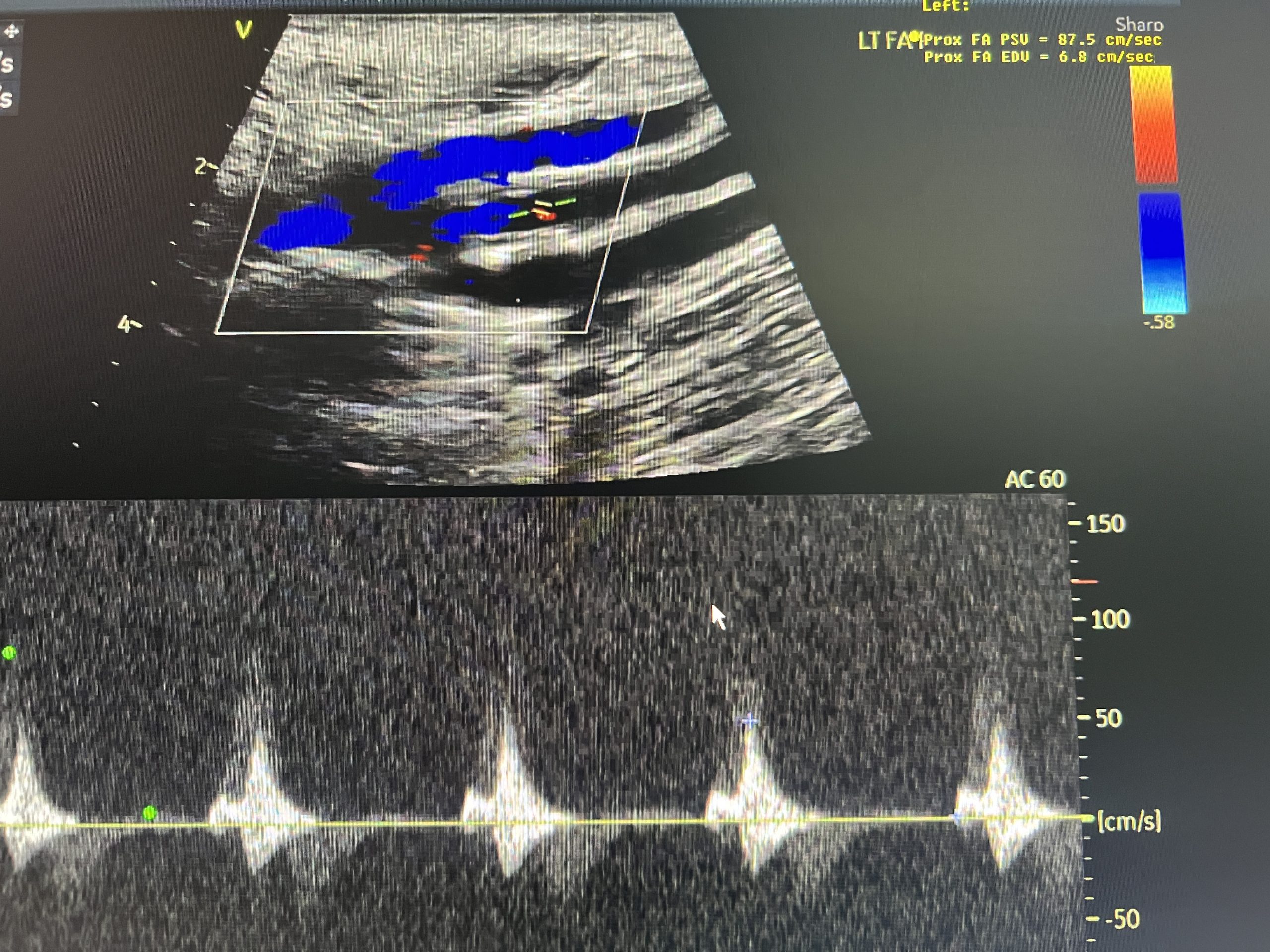
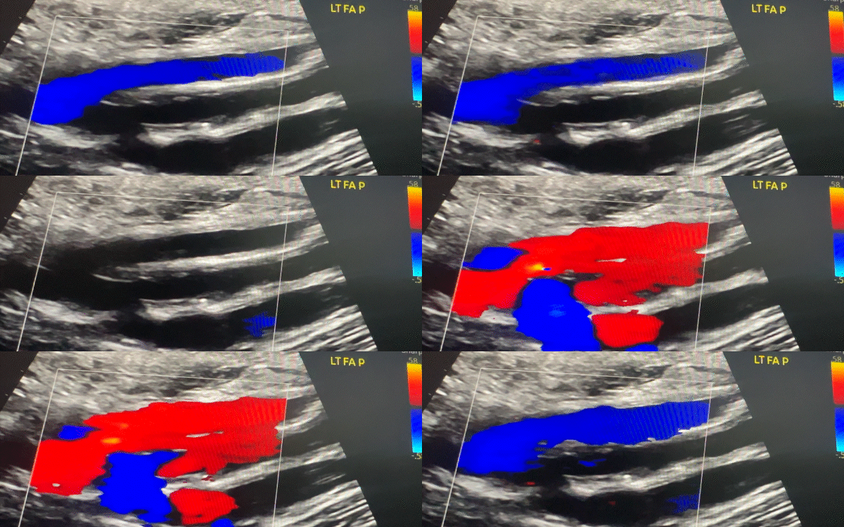
A patient presented with symptoms of vascular compromise in the left lower extremity—pain, coolness, discoloration, and cramping—prompting a referral for diagnostic imaging. An initial lower extremity arterial ultrasound revealed an occlusion of the distal left femoral artery, with reconstitution of blood flow at the level of the popliteal artery. These findings indicated a significant arterial blockage contributing to reduced circulation in the leg.
An endovascular procedure was promptly performed to place a stent and restore blood flow. The patient experienced temporary relief, but symptoms gradually returned. A follow-up CT angiogram confirmed that the stent had become occluded, signaling the need for surgical intervention. The patient subsequently underwent an autogenous bypass using the left great saphenous vein (GSV), connected from the common femoral artery to the distal popliteal artery to re-establish sustained blood flow to the limb.
 BB Imaging played a key role in providing detailed vascular imaging that continues to guide timely surgical decision-making. Moving forward, the patient will undergo annual imaging, including lower extremity arterial ultrasound, ankle-brachial indices (ABIs), and CT angiography (CTA), to monitor graft function and screen for new vascular disease. Ongoing care will include medication management and lifestyle modifications, coordinated through a multidisciplinary care team.
This case highlights the power of precision imaging in both acute diagnosis and long-term care. Through expert sonography and close clinical collaboration, BB Imaging helped support the patient on their path to surgical resolution, reinforcing the critical importance of advocating for proactive, informed care that can create better outcomes and preserve quality of life.
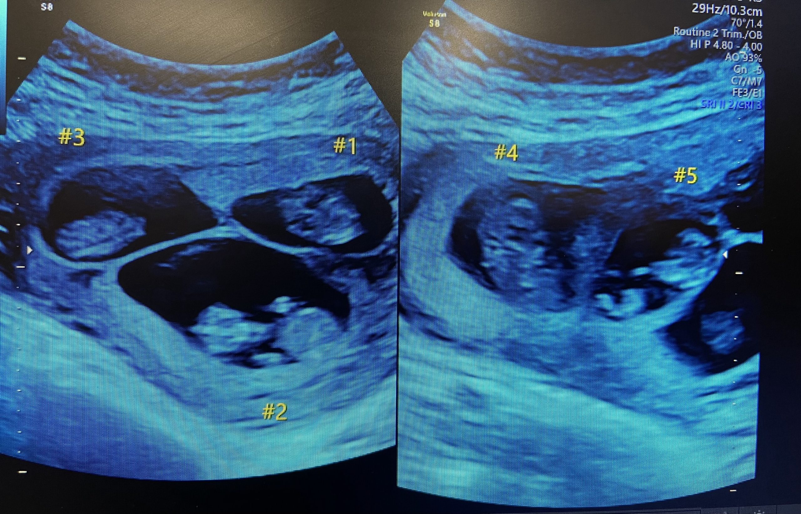
A 36-year-old patient was referred to maternal-fetal medicine for advanced imaging following confirmation of a quintuplet pregnancy—an occurrence so rare it affects roughly 1 in 55 million births. Her clinical profile included advanced maternal age, hypothyroidism, and a history of miscarriage.
The pregnancy followed a single round of intrauterine insemination (IUI). However, the patient reported taking emergency contraception after experiencing complications and being told she could not get pregnant at the time. She also disclosed a personal and family history of hyper-ovulation syndrome.
The fetuses, labeled 1 through 5, presented as triamniotic, with one monochorionic-diamniotic pair (3 and 4). The pregnancy included three female and two male fetuses. Routine imaging identified growth restrictions in 3, 4, and 5. Notably, 2 exhibited dolichocephaly, with a cephalic index of 64%, and 3 presented with severe intrauterine growth restriction (IUGR) and abnormal umbilical artery Doppler findings—including absent end-diastolic flow, an indicator of compromised placental blood flow.
Initially planned for delivery at 32 weeks, the pregnancy was expedited due to deteriorating conditions. At 28 weeks and two days, all five babies were delivered via Cesarean section. Despite low expectations for 3, who weighed just 500 grams, each baby survived, with none requiring intubation.
This case demanded extensive collaboration between BB Imaging and our physician partners. While our team supported the pregnancy with a series of critical scans, including fetal echocardiography, biophysical profiles, and growth monitoring, additional anatomical and growth assessments were conducted by our partner’s sonography team. It was a coordinated effort built on clinical trust, diagnostic precision, and a shared commitment to maternal-fetal health.
As one of the few documented successful quintuplet deliveries in El Paso, Texas, this case is a powerful reminder of what’s possible when physicians and sonographers work side by side to guide even the most complex pregnancies with precision and compassion.
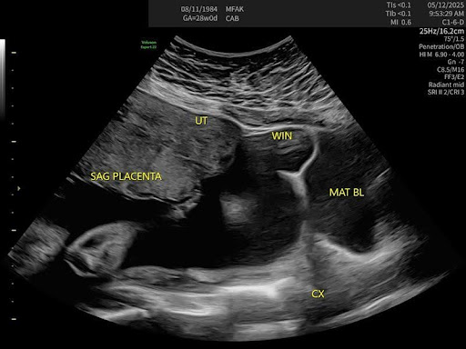
A 28-week pregnant patient (Gravida 8, Para 2) was receiving high-risk prenatal care due to several factors: advanced maternal age, chronic hypertension, obesity, and a history of two prior Cesarean deliveries (2020, 2022). During a routine visit, she reported localized pain in the lower uterine segment, right of midline, particularly when transitioning between sitting and standing.
Through a high degree of clinical coordination and further targeted ultrasound, the BB Imaging team supported the discovery of a uterine window defect—an area of thinning in the lower uterine segment. This structural vulnerability directly corresponded with the patient’s symptoms and raised concern for potential uterine rupture, especially given her obstetric history.
Following the findings, the patient was promptly sent to labor and delivery for administration of antenatal corticosteroids to accelerate fetal lung maturity, with a repeat Cesarean delivery planned within 24–48 hours. Thanks to early detection, the care team was able to proactively plan a carefully managed delivery, reducing the risk of future maternal and fetal complications. The critical findings were later confirmed at delivery via Cesarean section, reinforcing the importance of timely and informed obstetric intervention.
With highly detailed imaging and thoughtful clinical intuition, the BB Imaging team identified a subtle yet significant concern, reinforcing the value of timely, informed intervention in advancing care outcomes.

Ben Buentipo did not set out to start a company—he set out to solve a problem.
A sonographer by training, Ben had spent years honing his skills at the highest levels of obstetric practice, working across academic medical centers, large hospital systems, and private clinics. But over time, something kept catching his eye—and it was not just the images on the screen. It was the glaring inconsistency in sonography quality across the field.
In diagnostic ultrasound, image quality is not just about clarity. It is about outcomes. The accuracy of a diagnosis can hinge entirely on the precision and skill of the sonographer. That realization—how operator-dependent the entire process was—struck a nerve. Ben knew that too many patients were being let down, not because of the technology, but because of the talent gap.
Around that time, he and his wife, who had a background in business, started talking about launching something of their own. They tested a few ideas but ultimately returned to what Ben knew best: clinical excellence in imaging. More importantly, they started asking a fundamental question—how could they meet patients where they are?
While working at a high-acuity specialty clinic in the heart of a major city, Ben noticed something unsettling. Patients were driving over an hour for a single ultrasound appointment. They would take time off work, navigate traffic, sit through a medical visit, and drive another hour home. That was not just inconvenient—it was a barrier to care.
The answer became clear. What if they brought imaging to the patient, not the other way around? What if clinics in smaller towns or underserved areas did not have to turn patients away or send them across county lines? By embedding sonographers into local practices, BB Imaging could expand access, help clinics increase service capacity, and improve both outcomes and patient satisfaction.
From the beginning, the model had to work for everyone. It had to be better for patients—easier, more compassionate, more local. It had to be better for providers—reliable, high-quality, and revenue-positive. And yes, it had to work as a business. But at its core, BB Imaging was never just about filling staffing gaps. It was about protecting what matters most: timely, accurate care delivered by experts who treat every scan like someone’s future depends on it.
Because often, it does.
A 48-year-old male was referred for ultrasound evaluation after experiencing shortness of breath and atrial flutter—common symptoms with an uncommon cause. Imaging revealed heart failure secondary to biventricular non-compaction cardiomyopathy, a rare and often underdiagnosed condition affecting the heart’s muscle structure.
Management includes medications such as ACE inhibitors or ARBs, beta-blockers, diuretics, and devices like pacemakers, implantable cardioverter-defibrillators (ICDs), or cardiac resynchronization therapy (CRT). In severe cases, while a patient is awaiting a heart transplant, a ventricular assist device (R/L-VAD) may be required. Ongoing care emphasizes adopting a heart-healthy lifestyle and maintaining regular follow-ups with a dedicated care team.
This case underscores the value of high-quality ultrasound as a diagnostic tool and a clinical compass, guiding immediate and long-term care. By detecting a rare and potentially life-threatening condition, the BB Imaging team played a crucial role in enabling immediate case management, thereby reducing the risk of further complications and setting the stage for sustainable symptom and medication management.
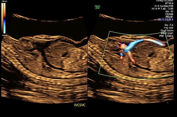
A patient was referred for a detailed anatomy survey due to Advanced Maternal Age. Upon ultrasound examination, ascites was initially suspected, but Doppler imaging revealed unexpected blood flow. Further evaluation confirmed an absent ductus venosus with the umbilical vein draining directly into the right atrium.
Additional findings included a single umbilical artery, persistent left superior vena cava, an apically offset mitral valve relative to the tricuspid valve, a duplicated right renal artery, and velamentous cord insertion. Due to the umbilical vein’s direct drainage into the right atrium, the perinatologist noted an increased risk for congestive heart failure. These findings were also associated with Noonan Syndrome.
An amniocentesis was performed and returned normal. A follow-up ultrasound later detected pericardial effusion and ventriculomegaly, both of which have since resolved. The patient remains under close monitoring until delivery.
Managing a clinic is a heavy lift, but partnership with BB Imaging can make the load a lot lighter. In this season of gratitude, let’s count the benefits that come from working together.
1. No-stress staffing
As the sonographer shortage continues, hiring struggles continue as well. On average, it takes between 36 and 42 days to fill a sonographer role, but, for rural locations, that timeline can extend to 180 days or more. And securing a start date isn’t the only challenge. Onboarding, training, and ramping up the schedule for a new employee takes approximately 12 weeks.
So, if we do the math, even in the best-case scenario, it will take clinics a minimum of 120 days before they have a fully productive sonographer working for them.
In contrast, BB Imaging can set up your clinic with highly skilled, knowledgeable sonographers in as little as 30 days. The sonographers we place in your facility are guaranteed to meet or exceed your requirements and are always graduates of CAAHEP-accredited programs with current registries.
So let BB Imaging take the stress out of staffing for your ultrasound department. We’ll handle all the recruitment efforts, cover the hiring costs, ensure credentials and training meet your requirements, and staff your facility in as little as 30 days with expert sonographers.
2. No-fuss schedule coordination
Because we’re staffing your facility with our sonographers, you also get to say “goodbye” to schedule management. We’ll always make sure your shifts are covered, regardless of our staff’s shifting needs, PTO requests, or unplanned leave.
We can even help cover for your staff sonographers. In 2024, we not only fulfilled 100% of our scheduled days—we also covered 93% of PRN requests from our partners.
3. Hands-free training and professional development
If you’ve worked with sonographers for any length of time, you know they’re passionate about growing their skillsets and leveling up their expertise. And while those qualities make sonographers a great hire, those expectations can put additional strain on employers who aren’t equipped to assist with training tools, practice time, and exam fees.
Because we’re entirely dedicated to high-quality sonography, BB Imaging has built a robust internal development program. We offer a variety of resources, assist with exam fees, and provide an elevating environment, with a lot of experts to learn from.
This means, first, that you can cross ‘training’ off your to-do list. Second, it means you get access to some of the most highly credentialed sonography experts in the nation.
For example, we did a little math using the ARDMS database. In the U.S., there are 125,000 total registrants, and only 3.4% are registered in fetal echocardiography. Contrast that to BB Imaging’s team, where 62% of our sonographers are fetal echo registered. Additionally, among our OB/GYN sonographers, 62% are also nuchal translucency credentialed and 62% are CLEAR credentialed.
4. Done-for-you benefits and payroll
A sonography shortage means a lot of competition. Many facilities compete for talent through strong benefits packages, higher pay, and sign-on bonuses. But offering these perks isn’t available to every facility.
Thankfully, you can hand all those tasks and costs over to BB Imaging. We offer sonographers a long list of industry-leading benefits—a list that makes us a destination workplace for the sonography community. And we’re always evaluating and adjusting sonographer salaries to make sure our pay rates are competitive.
All of these efforts result in low turnover. Sonographers who work for us want to keep working for us. In fact, our retention rates are always above 90%.
You don’t have to compete in a small talent pool, and the experts we provide will be around for a long time. Sounds like a win to us.
5. Easy scalability
BB Imaging can help you staff ultrasound departments in every kind of facility, from large hospitals to one-room clinics—and we’re happy to provide a single sonographer or bring the whole team.
Our contracts range from long-term partnerships to short-term needs to coverage for seasonal spikes, and we’re happy to adapt and grow with you as your patient demand fluctuates.
All of these options help our partners provide necessary appointments for their patients. Consistent coverage reduces the time until the next available appointment (from 8 weeks to 1 week for some!) and enables many of our partners to increase their services and make their ultrasound department more profitable.
6. Excellent diagnostics and patient care
Impressive credentials have to be followed up by highly accurate diagnostics and high-quality patient care, and our sonographers deliver on both counts.
We require 100% of our sonographers to scan to AIUM standards, even if the facilities they work in aren’t accredited. They’re also adept at onboarding provider preferences quickly and establishing them as part of our best practices and processes for your location. We also promise to care for your patients with the same level of compassion that you do, which leads to positive patient experiences and high patient satisfaction.
All these things work together to maintain and increase your reputation for outstanding ultrasound care.
7. Measurable cost savings
If you’re the kind of person who needs some numbers to better illustrate all the benefits of partnering with BB Imaging, we have you covered:
-
When BB Imaging handles sonographer recruiting, you save $10,500 in recruiting costs per open role.
-
A BB Imaging contract is based on usage. Compare that to an average salary of $80,000 per sonographer, plus thousands in benefits.
-
BB Imaging provides 100% of scheduled services. Compare that to the $22,000 in losses clinics incur for every week a sonographer is ill, injured, or on leave.
-
BB Imaging has created a destination workplace with a 90%+ retention rate. Compare that to a minimum cost of $130,000 for every sonographer turnover your clinic experiences.
Take the next step
You’ve counted the benefits. Are you ready to claim them now? We want to work with you. Just click the button below to set up a call.

