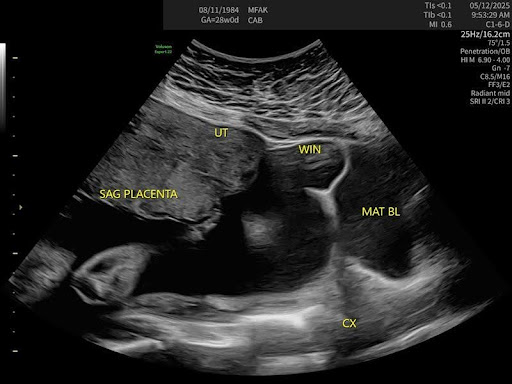
A 28-week pregnant patient (Gravida 8, Para 2) was receiving high-risk prenatal care due to several factors: advanced maternal age, chronic hypertension, obesity, and a history of two prior Cesarean deliveries (2020, 2022). During a routine visit, she reported localized pain in the lower uterine segment, right of midline, particularly when transitioning between sitting and standing.

Through a high degree of clinical coordination and further targeted ultrasound, the BB Imaging team supported the discovery of a uterine window defect—an area of thinning in the lower uterine segment. This structural vulnerability directly corresponded with the patient’s symptoms and raised concern for potential uterine rupture, especially given her obstetric history.

Following the findings, the patient was promptly sent to labor and delivery for administration of antenatal corticosteroids to accelerate fetal lung maturity, with a repeat Cesarean delivery planned within 24–48 hours. Thanks to early detection, the care team was able to proactively plan a carefully managed delivery, reducing the risk of future maternal and fetal complications. The critical findings were later confirmed at delivery via Cesarean section, reinforcing the importance of timely and informed obstetric intervention.

With highly detailed imaging and thoughtful clinical intuition, the BB Imaging team identified a subtle yet significant concern, reinforcing the value of timely, informed intervention in advancing care outcomes.

Ben Buentipo did not set out to start a company—he set out to solve a problem.

A sonographer by training, Ben had spent years honing his skills at the highest levels of obstetric practice, working across academic medical centers, large hospital systems, and private clinics. But over time, something kept catching his eye—and it was not just the images on the screen. It was the glaring inconsistency in sonography quality across the field.

In diagnostic ultrasound, image quality is not just about clarity. It is about outcomes. The accuracy of a diagnosis can hinge entirely on the precision and skill of the sonographer. That realization—how operator-dependent the entire process was—struck a nerve. Ben knew that too many patients were being let down, not because of the technology, but because of the talent gap.

Around that time, he and his wife, who had a background in business, started talking about launching something of their own. They tested a few ideas but ultimately returned to what Ben knew best: clinical excellence in imaging. More importantly, they started asking a fundamental question—how could they meet patients where they are?

While working at a high-acuity specialty clinic in the heart of a major city, Ben noticed something unsettling. Patients were driving over an hour for a single ultrasound appointment. They would take time off work, navigate traffic, sit through a medical visit, and drive another hour home. That was not just inconvenient—it was a barrier to care.

The answer became clear. What if they brought imaging to the patient, not the other way around? What if clinics in smaller towns or underserved areas did not have to turn patients away or send them across county lines? By embedding sonographers into local practices, BB Imaging could expand access, help clinics increase service capacity, and improve both outcomes and patient satisfaction.

From the beginning, the model had to work for everyone. It had to be better for patients—easier, more compassionate, more local. It had to be better for providers—reliable, high-quality, and revenue-positive. And yes, it had to work as a business. But at its core, BB Imaging was never just about filling staffing gaps. It was about protecting what matters most: timely, accurate care delivered by experts who treat every scan like someone’s future depends on it.

Because often, it does.

A 48-year-old male was referred for ultrasound evaluation after experiencing shortness of breath and atrial flutter—common symptoms with an uncommon cause. Imaging revealed heart failure secondary to biventricular non-compaction cardiomyopathy, a rare and often underdiagnosed condition affecting the heart’s muscle structure.

Management includes medications such as ACE inhibitors or ARBs, beta-blockers, diuretics, and devices like pacemakers, implantable cardioverter-defibrillators (ICDs), or cardiac resynchronization therapy (CRT). In severe cases, while a patient is awaiting a heart transplant, a ventricular assist device (R/L-VAD) may be required. Ongoing care emphasizes adopting a heart-healthy lifestyle and maintaining regular follow-ups with a dedicated care team.

This case underscores the value of high-quality ultrasound as a diagnostic tool and a clinical compass, guiding immediate and long-term care. By detecting a rare and potentially life-threatening condition, the BB Imaging team played a crucial role in enabling immediate case management, thereby reducing the risk of further complications and setting the stage for sustainable symptom and medication management.

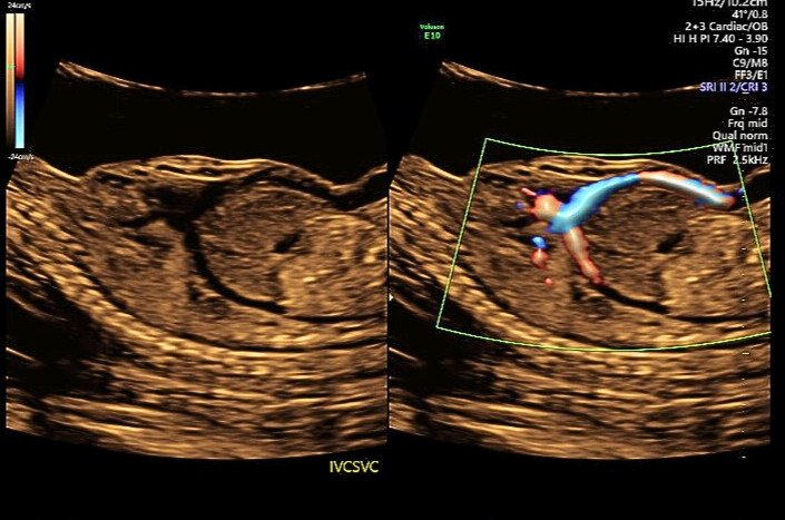
A patient was referred for a detailed anatomy survey due to Advanced Maternal Age. Upon ultrasound examination, ascites was initially suspected, but Doppler imaging revealed unexpected blood flow. Further evaluation confirmed an absent ductus venosus with the umbilical vein draining directly into the right atrium.

Additional findings included a single umbilical artery, persistent left superior vena cava, an apically offset mitral valve relative to the tricuspid valve, a duplicated right renal artery, and velamentous cord insertion. Due to the umbilical vein’s direct drainage into the right atrium, the perinatologist noted an increased risk for congestive heart failure. These findings were also associated with Noonan Syndrome.

An amniocentesis was performed and returned normal. A follow-up ultrasound later detected pericardial effusion and ventriculomegaly, both of which have since resolved. The patient remains under close monitoring until delivery.

Managing a clinic is a heavy lift, but partnership with BB Imaging can make the load a lot lighter. In this season of gratitude, let’s count the benefits that come from working together.

1. No-stress staffing

As the sonographer shortage continues, hiring struggles continue as well. On average, it takes between 36 and 42 days to fill a sonographer role, but, for rural locations, that timeline can extend to 180 days or more. And securing a start date isn’t the only challenge. Onboarding, training, and ramping up the schedule for a new employee takes approximately 12 weeks.

So, if we do the math, even in the best-case scenario, it will take clinics a minimum of 120 days before they have a fully productive sonographer working for them.

In contrast, BB Imaging can set up your clinic with highly skilled, knowledgeable sonographers in as little as 30 days. The sonographers we place in your facility are guaranteed to meet or exceed your requirements and are always graduates of CAAHEP-accredited programs with current registries.

So let BB Imaging take the stress out of staffing for your ultrasound department. We’ll handle all the recruitment efforts, cover the hiring costs, ensure credentials and training meet your requirements, and staff your facility in as little as 30 days with expert sonographers.

2. No-fuss schedule coordination

Because we’re staffing your facility with our sonographers, you also get to say “goodbye” to schedule management. We’ll always make sure your shifts are covered, regardless of our staff’s shifting needs, PTO requests, or unplanned leave.

We can even help cover for your staff sonographers. In 2024, we not only fulfilled 100% of our scheduled days—we also covered 93% of PRN requests from our partners.

3. Hands-free training and professional development

If you’ve worked with sonographers for any length of time, you know they’re passionate about growing their skillsets and leveling up their expertise. And while those qualities make sonographers a great hire, those expectations can put additional strain on employers who aren’t equipped to assist with training tools, practice time, and exam fees.

Because we’re entirely dedicated to high-quality sonography, BB Imaging has built a robust internal development program. We offer a variety of resources, assist with exam fees, and provide an elevating environment, with a lot of experts to learn from.

This means, first, that you can cross ‘training’ off your to-do list. Second, it means you get access to some of the most highly credentialed sonography experts in the nation.

For example, we did a little math using the ARDMS database. In the U.S., there are 125,000 total registrants, and only 3.4% are registered in fetal echocardiography. Contrast that to BB Imaging’s team, where 62% of our sonographers are fetal echo registered. Additionally, among our OB/GYN sonographers, 62% are also nuchal translucency credentialed and 62% are CLEAR credentialed.

4. Done-for-you benefits and payroll

A sonography shortage means a lot of competition. Many facilities compete for talent through strong benefits packages, higher pay, and sign-on bonuses. But offering these perks isn’t available to every facility.

Thankfully, you can hand all those tasks and costs over to BB Imaging. We offer sonographers a long list of industry-leading benefits—a list that makes us a destination workplace for the sonography community. And we’re always evaluating and adjusting sonographer salaries to make sure our pay rates are competitive.

All of these efforts result in low turnover. Sonographers who work for us want to keep working for us. In fact, our retention rates are always above 90%.

You don’t have to compete in a small talent pool, and the experts we provide will be around for a long time. Sounds like a win to us.

5. Easy scalability

BB Imaging can help you staff ultrasound departments in every kind of facility, from large hospitals to one-room clinics—and we’re happy to provide a single sonographer or bring the whole team.

Our contracts range from long-term partnerships to short-term needs to coverage for seasonal spikes, and we’re happy to adapt and grow with you as your patient demand fluctuates.

All of these options help our partners provide necessary appointments for their patients. Consistent coverage reduces the time until the next available appointment (from 8 weeks to 1 week for some!) and enables many of our partners to increase their services and make their ultrasound department more profitable.

6. Excellent diagnostics and patient care

Impressive credentials have to be followed up by highly accurate diagnostics and high-quality patient care, and our sonographers deliver on both counts.

We require 100% of our sonographers to scan to AIUM standards, even if the facilities they work in aren’t accredited. They’re also adept at onboarding provider preferences quickly and establishing them as part of our best practices and processes for your location. We also promise to care for your patients with the same level of compassion that you do, which leads to positive patient experiences and high patient satisfaction.

All these things work together to maintain and increase your reputation for outstanding ultrasound care.

7. Measurable cost savings

If you’re the kind of person who needs some numbers to better illustrate all the benefits of partnering with BB Imaging, we have you covered:

  1. When BB Imaging handles sonographer recruiting, you save $10,500 in recruiting costs per open role.

  2. A BB Imaging contract is based on usage. Compare that to an average salary of $80,000 per sonographer, plus thousands in benefits.

  3. BB Imaging provides 100% of scheduled services. Compare that to the $22,000 in losses clinics incur for every week a sonographer is ill, injured, or on leave.

  4. BB Imaging has created a destination workplace with a 90%+ retention rate. Compare that to a minimum cost of $130,000 for every sonographer turnover your clinic experiences.

Take the next step

You’ve counted the benefits. Are you ready to claim them now? We want to work with you. Just click the button below to set up a call.

 
Contact

If you’ve consumed any kind of media in the last six months, you’ve heard about how competitive the current job market is. Interestingly, the sonography industry appears to be largely sheltered from these trends, and the outlook for sonography jobs is much brighter than most careers.

The U.S. Bureau of Labor Statistics reports overall employment for ultrasound careers is expected to grow 10 percent from 2022 to 2032, with around 9,600 role openings each year. That’s a shocking number when you consider only 6,160 diagnostic medical sonography degrees were awarded in 2022.

Technological advances will likely seek to fill some of this gap, but sonographers will largely retain the luxury of choice as they consider where they want to work, who they want to work for, and the kind of pay and benefits packages they are willing to accept. And while the market in general will be less competitive than other careers, competition will be strong for the best roles.

Which is why you want an incredible resume.

Your resume is often your first impression with recruiters and hiring managers, so it needs to look its best, concisely convey your expertise, and answer the needs of the job description.

How to Make Your Resume Look Its Best

1 – Use a Template

We get it. You’re a sonographer, not a graphic designer. But that doesn’t mean you can’t have a gorgeous resume! Editable templates are available for free from companies like Adobe and Canva or can be purchased for a small cost on sites like Etsy and Creative Market.

P.S. We believe in this so much, we even made you a template just for sonographers! Click here to use our free Canva template.

2 – Use a Spell Checker

Making spelling mistakes (or using bad grammar and incorrect punctuation) is the fastest route to the digital discard pile. But it’s easy to save yourself from that result! Google Docs and Microsoft Word both feature built-in spell checkers and editors, or you can use a standalone tool like Grammarly. Many of these programs now also provide helpful, AI-powered suggestions to help you write clearly and concisely.

3 – Be Consistent

Help your readers get all the information they need by keeping design elements to a minimum. Don’t use bullet points, dashes, and numbers—just choose one. Use a minimal amount of font styles and formatting and keep your color scheme minimal too. In the case of a resume, the design should enhance the copy you’ve written, not compete with it.

P.S. All of these rules get much simpler when you choose a ready-made template! 

4 – Export as a PDF

Exporting your resume as a PDF helps it maintain its formatting so that it appears correctly on other screens. The last thing you want is to create a stunning resume and then have it show up as a scrambled mess on the recruiter’s computer. Here’s how to take this crucial last step in three widely used programs:

  • Microsoft Word: Click File, click Save As, then click Download as PDF

  • Google Docs: Click File, click Download, then click PDF Document

  • Canva: Click Share, Click Download, Click the File Type dropdown arrow, Click PDF

How to Convey Your Expertise

5 – Write Clearly and Concisely

If you want your experience to shine, you can’t bury it in long, complex sentences. Instead, use short, snappy sentences that begin with a verb (action word). Here are a few examples:

  • Inspected and reviewed patient charts, labs, and medical history

  • Performed 8-10 sonographic examinations per day

  • Evaluated sonograms to identify or rule out patient/fetal anomalies

  • Created reports to aid physicians with documentation and diagnosis

6 – Include Transferable Skills

Don’t neglect to include work experience just because it isn’t in the medical field. Sonography requires many skills that transfer well between industries, and those skills could be the deciding factor between you and another candidate. Worked in retail? Mention skills like customer communication or restocking supplies. Spent some time in customer service? Highlight your interpersonal skills and problem-solving abilities. Work a side hustle? Draw attention to your ability to take the initiative and handle all the details.

7 – Amplify Your Achievements

Don’t use your work experience section to list all your daily activities. Instead, think about it like a highlight reel. This section should flow seamlessly from your most to least recent roles and hit all your major accomplishments for each role. Did you give a presentation or publish a paper? Were you considered a subject matter expert for a particular exam? Did you create helpful resources or establish more effective processes? These are all high-level contributions. Draw the most attention to those items.

8 – Tell Your Story

A touch of personality can go a long way, so don’t be afraid to tell more of your story. Try including a brief summary of what made you interested in sonography or why you’re passionate about patient care. And if you have some room, try including your interests, group affiliations, or volunteer work. The goal of these sections is to be relatable and memorable.

9 – Prep Your References

If you’re sending out your resume, you should also have your references prepped. The general rule of thumb is to provide three references, usually upon request. It’s best for these to be professional or educational contacts rather than personal or character references.

But… before you list someone as your reference, get their permission. Unexpected calls are often ignored. Worse, the call could reflect poorly on you if the reference mentions they aren’t prepared. Asking their permission also gives you the opportunity to update their contact information, check their availability, and let them know what position you’re interviewing for.

Once you have their permission, make sure you have included the following items for each reference listed:

  • Their first and last name

  • Their current job title and company

  • Their direct phone number and email address

  • Your relationship to the reference (professor, team lead, direct supervisor, etc.)

And when you land the job, remember to thank them. Even in our technologically advanced age, nothing beats a hand-written thank you card.

Bonus Tip: If you’re a new grad looking to stand out, consider sending preceptor letters with your resume. This is a great way to stand out—even if you’re just starting out!

How to Answer the Job Description

10 – Include Keywords 

Think about a job description like a series of questions. Can you do the tasks required for this role? Do you have the requisite education and credentials? Your resume is your answer to these questions—and repeating their words and phrases back to them is one of the easiest ways to provide a clear answer.

Maybe they use the term “ultrasound tech” rather than “sonographer” and “procedure” rather than “exam.” Make a quick edit to adopt their terminology. Review their big-picture description of the role. What pieces can you add to your summary or objective statement? Scan the list of specific tasks listed. Can you edit or add anything to your work experience section that mirrors their language? These changes are small, but they can have a big impact.

11 – Use Numbers

On a page full of text, numbers tend to stick out. Look at the examples below:

  • Reviewed fetal images to find or rule out anomalies

  • Collaborated with 8 perinatologists and 3 pediatric radiologists to review fetal pathology

  • Served as lead sonographer on clinical team

  • Led a team of 7 sonographers and 2 rotating interns

  • Presented at several industry events

  • Gave 5 industry presentations in a 2-year timespan

See what we mean? Numbers are concrete pieces of data that showcase the degree of your capability—and they are powerful.

12 – Update Your Contact Information

Provide a professional email address, make sure your phone number is current, and make sure they are accurate. It would be a shame to miss out on an opportunity because you listed your contact information incorrectly.

Speaking of missing out… email providers are notorious for sliding recruiter emails straight into your junk folder. If you’re actively applying, remember to check your junk or spam folders often for miscategorized emails. If you want to be proactive, add a company’s email domain to your safe senders list after applying so you never miss a message. And even better yet, opt into text messaging so you’re sure to see every message quickly and conveniently.

13 – How to Nail the Interview

Resumes are only the beginning of the job hunting process, but don’t worry, we’ve got your back when it comes to successful interviews too! Check out these resources:

There you have it! With these eleven tips in mind, you should be well on your way to a stellar sonography resume. Don’t forget to use our free sonographer resume template, and when it’s ready, be sure to stop by our job board.

 
Discover Careers

At BB Imaging, the health of our sonographers is directly related to the health of our company, our patients, and our providers’ clinics. So, we’ve made it our mission to ensure these valuable professionals stay healthy in both body and mind.

What follows is a helpful guide for:

  • Understanding fatigue at work

  • Identifying the signs and symptoms

  • Preventing burnout and advocating for resources

  • Seeking professional treatment

Defining Work-Related Fatigue

What is fatigue? Most people know they are fatigued when they feel tired, weary, or sleepy. This feeling usually follows prolonged mental or physical activity, loss of sleep, or extended periods of stress. While this understanding of fatigue is true, it’s far from comprehensive.

Fatigue is a highly complex phenomenon, with a wide range of symptoms and causes. It can occur at the individual, interpersonal, or work level—or all three. And sometimes, it can be hard to separate the symptoms and causes into neat buckets, because work fatigue is influenced by personal and interpersonal situations, and vice-versa.

A related term to fatigue we’re all familiar with is “burnout.” Formally known as occupational burnout syndrome, burnout gives us a word to use when specifically discussing work-related stress that isn’t being successfully mediated.

Burnout in Healthcare

You don’t have to scroll through news sites for long to realize that burnout rates in the clinical environment are skyrocketing. You probably don’t have to go to news sites at all—you’ve likely seen, heard, and felt it in your own workplace.

Burnout in healthcare can be especially problematic because of its impact, not just on the individual experiencing burnout, but also on others. Burnout has been shown to:

  • Increase the risk of work-related accidents

  • Increase practitioner injury

  • Result in poorer patient outcomes

  • Result in longer inpatient stays

  • Slow healthcare team reaction times

  • Increase error rates

Burnout in Sonography

Among sonographers specifically, fatigue can cause missed pathology, misinterpretation of images, a reduced level of patient care, and poor interpersonal skills.

This is why keeping sonographers from feeling fatigued is so important. Unfortunately, the industry’s efforts in this area haven’t been especially successful on a broad scale.

A 2022 study examining the causes of sonographer burnout found two main areas for improvement: workload and circumstances related to adverse news. Other studies back up these concerns. Cohen et al found that increased workloads led to physical symptoms including eye strain, muscle aches and pains, carpal tunnel syndrome, and (you guessed it) fatigue. In addition, a 2018 study by Johnson et al found that delivering adverse news contributes to anxiety and depression.

The ultimate impact of sonographer burnout is made clear in a study released earlier this year by My Tran, MS, RDMS, RVT.

When asked to provide the likelihood of leaving the profession as a sonographer…

  • 24.3% answered strongly agree

  • 43.8% answered agree

That means nearly 70% of survey respondents expect to leave the sonography industry rather than making it a life-long career.

Again, when asked if they were currently considering leaving the profession…

  • 16.2% strongly agreed

  • 28.9% agreed

That means more than half of those who expect to leave the industry are currently considering a transition to another profession. This may go without saying, but if those numbers hold, we’re looking at a dire scarcity of ultrasound services.

Symptoms and Signs of Burnout

Because it is complex, burnout has a variety of symptoms and can manifest itself differently in different people. We’ve compiled a list of common symptoms below, but it isn’t exhaustive. See if any sound familiar:

Personal

  • You don’t have energy for leisure activities

  • You’re experiencing eye strain or muscle aches and pains

  • You’re experiencing headaches or stomach problems with no known cause

  • You’re using food, drugs, or alcohol to feel better or numb how you feel

Interpersonal

  • You feel removed from the people you work with

  • You lose patience with co-workers

  • You find it difficult to communicate compassionately with patients

Workplace

  • Your working hours and overtime have increased

  • You’re performing an increased number of scans per day or week

  • Your responsibilities don’t align with your capacity

  • You’re experiencing an increase in exposure to stressful situations

  • Your productivity or task capacity is greatly reduced

  • You feel tired before arriving at work

  • You feel mentally or emotionally drained during work

  • You feel worn out or weary after work

  • You’re questioning the value of your work

  • You feel cynical toward your occupation and removed from your work

  • You find it difficult to focus on the task at hand

  • You feel little to no satisfaction from your accomplishments

  • You feel let down by your job

  • You’re doubting your skills and abilities

Preventing and Treating Burnout

Now that everyone is on the same page regarding the seriousness of this issue, let’s see what we can do to curb that trend and help our nation’s sonographers experience burnout-free work lives.

On a personal level, sonographers have a lot of options when it comes to preventing feelings of burnout:

  • Eating a nutritious diet

  • Exercising regularly (bonus points if you go outside!)

  • Practicing good sleep hygiene

  • Engaging in a stress-reduction activity like deep breathing, yoga, or meditation

  • Starting a regular self-reflection practice

  • Caring for your body while you’re at work by practicing healthy ergonomics

  • Taking time off before burnout sets in

  • If PTO isn’t an option, try taking a break without using PTO 

On an interpersonal level, sonographers (like everyone else!) need community. Sometimes talking about issues helps us to release the stress associated with those issues. Try these burnout-relieving conversations:

  • Talk to peer sonographers

  • Talk to other healthcare team members

  • Form a support group, even if you can only meet for a few minutes each day

  • Connect with a friend or family member

Now we come to the workplace. The work environment can feel out of our control, but there are usually some things you can do to advocate for improvements:

  • Reach out to your supervisor and request decreased hours or workload

  • Ask the human resources department for employee resources

  • Participate in organizational initiatives where you can make your voice heard

  • Advocate for mental health benefits and mindfulness training 

  • Ask for and assist in setting protocols for delivering adverse news

Seeking Professional Help

Don’t skip this section! We know it can be difficult to ask for help, especially as a professional caregiver, but we all need help sometimes. Asking for help is a sign of strength, not of weakness.

Here are a few signs that it’s time to be strong and ask for help:

  • You experience symptoms of anxiety including an increased heart rate, difficulty breathing, and feelings of nervousness, even outside of stressful situations

  • You regularly experience symptoms of depression including social withdrawal, poor sleeping, weight changes, or aches and pains

  • You experience feelings of sadness or guilt and a sense of loss regarding formerly enjoyed activities

  • You experience feelings of apathy and hopelessness or thoughts of suicide

Seriously, don’t skip over this. If the cost of seeking professional help is a barrier, ask your HR representative about mental health benefits – you may have free access to resources. If you’re not sure where to go, see this list of top-rated online therapy options—many have coupon codes available.

What would you add? Are there symptoms we missed? Do you have suggestions for prevention and treatment? Let us (and the sonographer community) know in the comments!

Please note: Information in this blog post should not replace the advice of a physician or mental health expert.

Federally Qualified Healthcare Centers (FQHCs) play a major role in improving maternal outcomes by delivering care to low-income, rural, and minority groups. According to the Health Resources & Services Administration, more than 12,000 FQHC sites across the country cared for more than 28.6 million patients in 2020 alone. That includes nearly one‐third of low‐income women of reproductive age and 8% of all pregnant patients nationwide.

In recent years, our nation’s reliance on these centers has grown. A 2023 community health center report found 6 million more patients are visiting FQHCs, accounting for a 24% increase since 2015. This rise in dependency is mirrored by the 186 rural hospital closings between 2005 and 2022. Counties that experienced a hospital closure became more economically challenged, were more likely to experience primary and dental care shortages, and had a greater number of non-White residents.

FQHCs fill the care gap for these patients. But as other care locations continue to decrease, FQHC leaders are looking to telemedicine and technological innovation to help them continue to offer desperately needed, high-quality care.

How a Remote Ultrasound Solution Can Help

That’s where teleultrasound® enters the equation. Relying on the expertise of an experienced perinatal telesonographer, our prenatal ultrasound solution, TeleScan, enables FQHCs in any location to offer in-house ultrasound.

In brief, here’s how it works:

  1. A designated healthcare worker learns to capture cine clips and upload them using an ultrasound machine and the TeleScan software.

  2. Once uploaded, the cine clips are reviewed by a certified perinatal telesonographer who provides annotations and measurements and compiles a preliminary diagnostic report.

  3. When the report is ready, the provider accesses the report through the TeleScan software. They can edit, add notes, and sign the report.

  4. Reports are turned around in about 30 minutes, enabling the provider to relay results while the patient is still in-clinic.

  5. Patients can also opt to have selected keepsake images sent to their phone.

By leveraging remote expertise, FQHCs realize four important benefits. Adding a telesonographer to the team…

1. Addresses the Sonographer Shortage

Hiring and retaining sonographers is increasingly difficult, with underserved and rural locations often unable to compete for top talent, and amid a sonographer shortage, the competition is fierce. From 2011 to 2021, the demand for ultrasound exams increased from 38.6 million to 59.8 million, accounting for a 55% jump. In that same time, however, the number of available sonographers increased by only 44%. To put it in starker terms, diagnostic medical sonography programs across the nation graduated 5,393 sonographers during those ten years, but they couldn’t come close to filling the 25,162 sonographer positions available.

As demand continues to outpace supply by large margins, it’s become clear that we need technology to help fill the gap and extend the reach of these invaluable experts. Adding TeleScan as an ultrasound partner provides FQHCs with access to an entire bench of telesonographers with deep expertise and experience in maternal-fetal ultrasound. These professionals:

  • Graduated a CAAHEP-accredited DMS program

  • Hold active registries in OB/GYN and Fetal Echocardiography through the ARDMS

  • Hold active Nuchal Translucency, Nasal Bone, and Cervical Length Education and Review credentials

  • Have at least two years of maternal-fetal medicine sonography experience

To the point: Adding a telesonographer to the team allows FQHCs to overcome the sonographer shortage and bypass the competition for talent.

2. Reduces Barriers and Enhances Accessibility

Many factors work against positive health outcomes in underserved communities, and they often come in the form of social and systemic barriers. Rurality, medical disenfranchisement, and social determinants of health (SDOH) like childcare, transportation, and financial constraints can all impede the provision of high-quality healthcare.

A recent study in northern Canada discovered geographic isolation from imaging facilities was an especially significant barrier for patients trying to access ultrasound imaging. Additional barriers presented themselves when travel for ultrasound became necessary, including fear of travel, isolation, financial challenges, and unfamiliarity with large cities.

To prevent undue stress and discomfort for patients, it makes sense to bring prenatal ultrasound technology to them. By enabling in-house diagnostic ultrasound, FQHCs can minimize distance to care as well as the negative side effects patients experience from travel.

Our own data shows a telemedicine approach is effective. An FQHC in Texas surveyed its patients and discovered 100% of them reported being likely to show for an appointment if ultrasound was offered. Many were true to their word, and patient compliance increased from the 70th percentile to 86% in the first three months following the implementation of TeleScan.

To the point: Adding a telesonographer to the team enables FQHCs to offer in-house ultrasound services, prioritize patient comfort, and increase appointment adherence.

3. Adds Operational Efficiency and Profitability

Non-local ultrasound appointments aren’t just hard on patients—they can often be time-consuming and chaotic for FQHCs to manage too. Working with third-party imaging centers puts FQHC staff in a middle-man role, scheduling and rescheduling appointments, chasing down reports for days (if not weeks), and laboring through a cumbersome and disjointed workflow.

In contrast, TeleScan provides a single end-to-end solution that puts FQHCs in charge of the entire patient experience. Scheduling and exams are performed in-house, and reports are turned around in 30 minutes, leaving plenty of time for a provider consultation while the patient is still in the clinic. Quicker and smoother exams also reduce appointment wait times, often by 50-75%.

Efficiency and an increase in patient compliance can also impact an FQHC’s profitability. TeleScan’s handy return on investment calculator estimates the level of ROI a facility could expect based on its billing structure. This is just an estimate, and revenues could increase substantially when services are scaled up or added to satellite clinic locations.

To the point: Adding a telesonographer to the team increases efficiency and could yield a positive return on investment.

4. Confronts Maternal Health Statistics

Everyone has seen the statistics released by the March of Dimes last year, which were well circulated by the news media. The report showed maternity care deserts are home to more than 2 million women and an additional 3.5 women live in counties with limited maternity care access. Rural and minority populations are disproportionately affected.

Slowly but surely, telemedicine is working to reverse these trends and improve outcomes in women’s health. TeleScan stands to make an important contribution to this effort by identifying high-risk pregnancies early and giving FQHC providers time to treat, monitor, and plan for a healthy delivery. That’s why telesonographers are equipped to analyze ultrasounds during all trimesters of pregnancy, including anatomy scans, growth scans, nuchal translucency exams, and biophysical profiles.

To the point: Adding a telesonographer to the team extends accurate diagnostics and maternal care to low- and no-access counties.

How to Add a Telesonographer to Your Team

FQHCs have a deep understanding of the challenges surrounding maternity care for underserved patient populations. Sonographers are difficult to hire, patients experience a variety of barriers to care, clinical operations can get messy, and maternal health statistics are disheartening.

But telesonography provides a truly effective ultrasound solution for FQHCs, and adding a telesonographer to the team can provide a host of benefits for providers, patients, and facilities alike.

 
Add a telesonographer to your team

Lately, the medical community has been abuzz with an intriguing observation: a noticeable uptick in emergency cerclage procedures, particularly among women who were once considered low-risk.

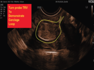
Emergency cerclage, a critical intervention for pregnant women facing cervical insufficiency, helps prevent the risk of premature delivery by reinforcing the cervix. This condition, where the cervix begins to prematurely dilate and thin out, can lead to preterm labor—a serious threat to both mother and baby. By keeping the cervix securely closed until the baby reaches full term, emergency cerclage is essential for minimizing the complications associated with premature births.

So, what exactly is a cerclage, and how do doctors determine who needs one? Let’s dive in and explore more about this vital procedure and its implications.

Historical Context and Evolution

The practice of cerclage dates back to the 1950s when doctors first began using stitches to support high-risk pregnancies. Over the decades, advancements in medical technology and surgical techniques have significantly enhanced the safety and effectiveness of cerclage procedures. Innovations such as precision suturing tools and improved imaging technology enable physicians to perform these procedures with greater accuracy and reduce complications.

Types of Emergency Cerclage Procedures

There are three primary types of emergency cerclage procedures in use today:

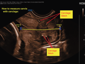
  • McDonald Cerclage: The McDonald cerclage is the most common procedure. It involves placing a stitch around the cervix, essentially creating a supportive loop that holds the cervix closed.

  • Shirodkar Cerclage: The Shirodkar cerclage employs a permanent suture in the cervix. Unlike the McDonald cerclage, this method uses more complex suturing techniques to ensure the cervix remains closed.

  • Abdominal Cerclage: An abdominal cerclage requires more invasive abdominal surgery. This procedure is often reserved for women who have experienced failed cerclages previously or have anatomical challenges that make other types of cerclage less effective.

Increasing Awareness and Screening

In recent years, awareness of cervical insufficiency has significantly increased. New strategies are being developed to enhance the accuracy of cervical length assessments by focusing screenings on low-risk women who have identifiable risk factors for a short cervix. These risk factors include minority race, tobacco use, prior preterm birth, and prior cervical procedures.

Additionally, BMI could be considered a potential risk factor for a short cervix in future screening algorithms. Both obesity and cervical insufficiency are major contributors to pregnancy-related morbidity. In the USA, over 20% of women of reproductive age are obese (BMI > 30), a figure that is steadily rising.

When is Cerclage Recommended?

Cerclage is recommended for women with a history of cervical insufficiency, multiple preterm births, or specific changes in the cervix observed via ultrasound. In more detail, it’s typically advised for women who’ve experienced 3+ preterm deliveries or mid-trimester losses, making a history-indicated cerclage vitally important in such cases.

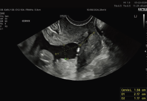
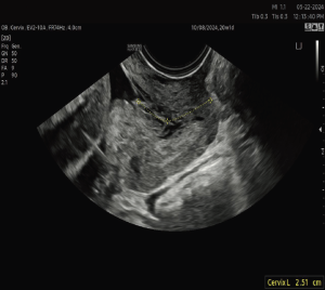
An ultrasound-indicated cerclage is a wise choice when a woman’s cervical length measures less than 24 mm, particularly if she has a history of spontaneous preterm births or mid-trimester losses. Significantly, for those expecting twins, the threshold for intervention is even shorter, with cervical lengths under 15 mm suggesting the need for this supportive measure.

Generally, cerclage placement is considered before the 24-week mark, offering a promising avenue for maintaining a healthy pregnancy and bringing new hope and optimism to expecting mothers.

The Procedure and Its Risks

Typically performed under anesthesia in an outpatient setting, cerclage is generally considered safe. However, the procedure does carry some risks, including infection, bleeding, preterm labor, and membrane rupture. Post-cerclage, some cramping or spotting is normal, but many women find they can swiftly return to their usual routines, embracing a healthier pregnancy journey ahead.

Alternatives to Cerclage

For patients who opt out of a cerclage or have a cervical measurement that is borderline short at over 25mm, there are several promising alternatives to consider. These options include bed rest, which can reduce physical stress on the cervix, and progesterone therapy, often used to help maintain pregnancy by supporting the cervical structure. Cervical pessaries, small silicone devices placed around the cervix, can also provide extra support and may prevent premature shortening.

Regular cervical length monitoring is another vital aspect of managing this condition, ensuring any changes are promptly detected. It’s crucial to have an in-depth discussion with healthcare providers about these treatments, enabling patients to make the most informed and personalized decisions for their health and their baby’s well-being.

Post-Procedure Care and Removal

Once placed, the cerclage typically remains in situ until around the 36th to 37th week of gestation. At this point, the obstetrician usually removes it. The removal process is generally quick and straightforward, often not requiring anesthesia. This helps ensure a smooth and stress-free experience for our patients as they approach the final weeks of their pregnancy.

Why It Matters to Sonographers

The increase in emergency cerclage procedures among low-risk patients highlights the essential role of skilled ultrasound technicians in modern obstetric care. Our expertise in imaging and assessment enables early detection, timely interventions, and provides critical support to expectant mothers facing anxiety. This precision fosters trust, improves collaborative decision-making, and enhances maternal and fetal outcomes. As challenges evolve, we must stay committed to excellence, compassion, and innovation to ensure optimal care for all expectant mothers.

References:

https://obgyn.onlinelibrary.wiley.com/doi/full/10.1111/aogs.13881

https://karger.com/goi/article/87/5/299/823900/Weekly-Differences-in-the-Prevalence-of-a-Short

https://www.medicalnewstoday.com/articles/short-cervix#outlook

https://emedicine.medscape.com/article/1848163-overview?form=fpf#a1 

About Lashell: Lashell Jones, RDMS, RDCS (FE) is a highly skilled ultrasound specialist with more than 20 years of experience. She currently holds the Lead Sonographer position at Maternal Fetal Associates in Reston, Virginia. Lashell is also an active participant in the Inteleos Diversity and Inclusion task force, an author of PREPRY study resources, and a registry review tutor. She is on a mission to improve medical awareness and education within the ultrasound industry.

One look at industry headlines is all you need to realize artificial intelligence (AI) is improving at breakneck speed. And while we’re big proponents of tech-enabled solutions that increase access to ultrasound care, we’re also convinced that AI, in its current state, isn’t ready to go it alone just yet.

Even if you just invested in the fanciest, AI-enabled ultrasound technology on the market, here are some reasons you still need a sonographer to oversee it:

1. Accuracy Verification

AI, while powerful, is not infallible. We don’t mean to be dramatic, but an inaccurate diagnostic report can mean the difference between treatment and suffering—or life and death—and it’s our responsibility to provide the greatest accuracy to every patient. Today, the greatest chance for accuracy usually doesn’t reside with AI-only analysis.

For example…

Consider this study regarding AI and physician assessment of lung ultrasound to identify pulmonary edema. The physician was 96.7% sensitive and 79.1% specific compared to the AI software’s 95.6% sensitivity and 64.1% specificity. This demonstrates that AI was on par when it came to detecting pulmonary edema but returned significantly more false positives than the physician.

This demonstrates the need for a human counterpart to verify the accuracy of AI-generated findings. Diagnostic accuracy is crucial to achieving correct diagnosis, administering helpful treatment, and ultimately, achieving a healthful outcome.

2. Anomaly Detection

AI algorithms are trained to detect anomalies based on the data they have been trained on—data that can be low-quality and is often incomplete. Sonographers need to review AI’s work to catch anomalies that don’t fit within the parameters set by the training data to ensure that rare or unusual cases are not overlooked.

For example…

In a recent study regarding AI in fetal ultrasound, the authors extolled AI’s ability to identify prenatal ultrasound views, assist in gestational age estimations, and measure biological parameters. These are helpful capabilities, but when it came time to discuss limitations, the authors admitted, “Most data sets lack pathological cases and only include healthy fetuses and pregnant women. The uneven training data sets result in poor algorithm training performance.”

Until datasets are both high-quality and complete, ultrasound images need to be reviewed by sonographers for pathology detection. AI will also have to lean on them for high-quality training images—and lots of them—to increase its ability to identify anomalies.

3. Complex Decision-Making

While AI has been proven to aid decision-making processes, it often suffers from the ‘black box problem’—the inability to show how it came to its conclusion. Because AI often can’t explain its outputs, an expert sonographer who can knowledgeably relay their findings is a highly valuable asset to any care team.

For example…

In their 2023 article, Responsible application of artificial intelligence in health care, Obasa and Palk conclude, “Black box systems raise numerous ethical concerns, including explicability […] When there is a high risk of harm or negative outcomes associated with the decisions of such systems, we should be able to ascertain a full understanding of the decision-making process of the system. This implies that black box systems, for which such an explanation is not possible, should not be used with procedures that carry such high risk.”

As noted, explainability is paramount to making informed decisions. Understanding the how’s and why’s of a patient’s case, and being able to communicate them to the patient, is key. AI’s inability to answer these questions leads to a continued dependence on sonographers and providers to complete these tasks.

4. Ethical Responsibility

Ultimately, sonographers and other healthcare professionals bear an ethical (and sometimes legal) responsibility for patient care. But understanding AI’s level of responsibility in a care setting is an area of concern with more questions than answers. Since current laws regulating ethical and legal responsibility didn’t see AI coming, human oversight of AI is the best, and safest, course of action.

For example…

Adding AI into the care equation can make it difficult to attribute responsibility to just one party. This is known as diffusion of responsibility. This 2022 article identified three levels of responsibility diffusion that occur with the use of AI in a clinical context:

  1. Causal: responsibility for harm sustained from misdiagnosis or misguided treatment

  2. Moral: responsibility to prevent harm and promote welfare (think the Hippocratic oath)

  3. Legal: responsibility to accept liability and to compensate for harm done

It’s currently difficult to prove AI should be held to any of these levels of responsibility. So, while it doesn’t solve all the problems of responsibility diffusion, reviewing AI’s work is still an important step in providing ethical patient care.

Yes, and…

So far, our discussion highlights the importance of human oversight for AI in medical imaging. A real-life sonographer is still needed if we’re going to provide accurate, ethical, patient-centered care. But we want to say more on this issue: Yes, a sonographer is still needed… and one of the greatest tools you can provide them is AI.

For example…

  • A deep learning-based graph output helped sonographers improve fetal cardiac ultrasound screenings for more accurate detection of congenital heart disease.

  • Automated segmentation of images reduced keystrokes by 80% for fetal brain exams and reduced delineation of organs at risk to just 20 minutes (compared to 1-3 hours when completed manually).

  • AI automations in fetal ultrasound acquired and measured images, enabling sonographers to concentrate better on image interpretation.

  • Automated quality scores for fetal cardiac four-chamber views were a useful training tool to improve scanning and provide more consistent images.

Considering the limitations listed above, and the examples provided here, we don’t think the AI conversation pits sonographers against emerging technology. Rather, we think emerging technologies enhance and expand their already incredible skillset.

Have we convinced you? Do you agree that sonographers are still needed—and that AI is a helpful tool? Let us know in the comments.