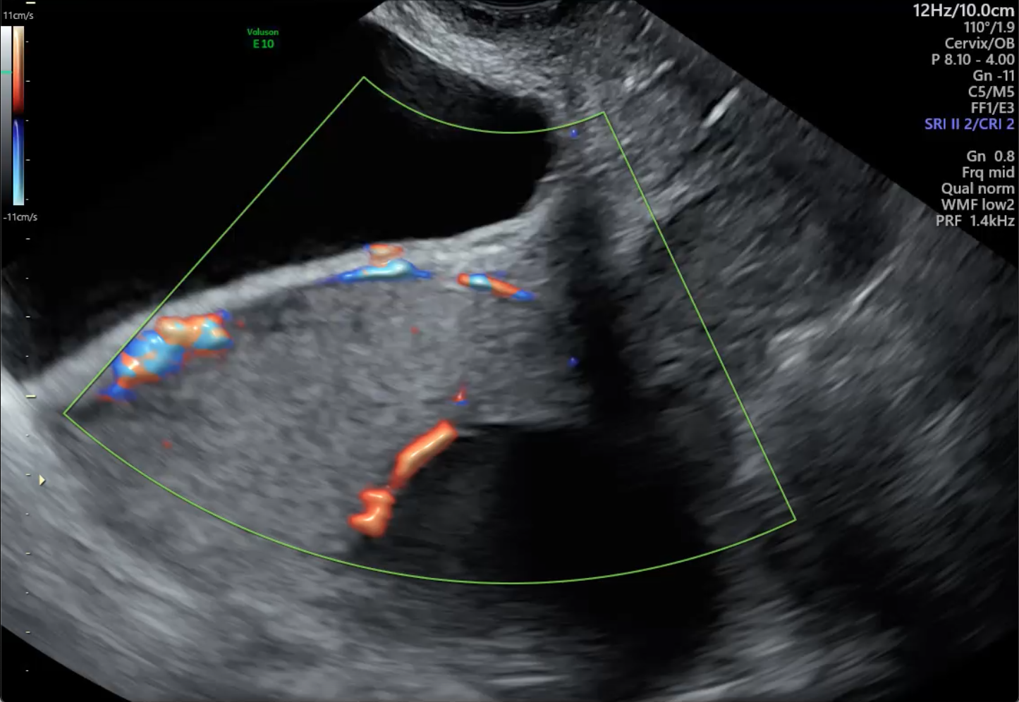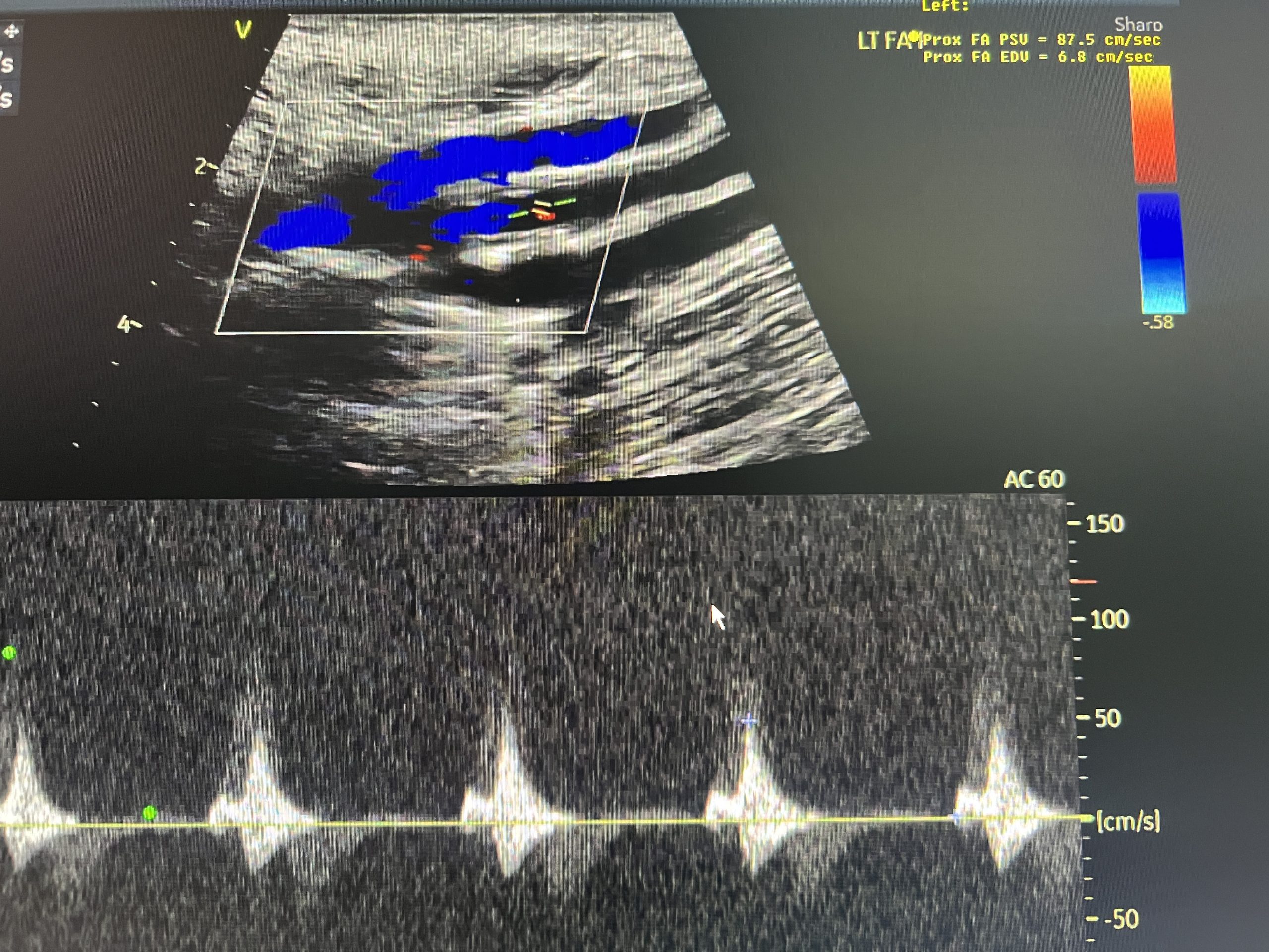
A patient presented with symptoms of vascular compromise in the left lower extremity—pain, coolness, discoloration, and cramping—prompting a referral for diagnostic imaging. An initial lower extremity arterial ultrasound revealed an occlusion of the distal left femoral artery, with reconstitution of blood flow at the level of the popliteal artery. These findings indicated a significant arterial blockage contributing to reduced circulation in the leg.
An endovascular procedure was promptly performed to place a stent and restore blood flow. The patient experienced temporary relief, but symptoms gradually returned. A follow-up CT angiogram confirmed that the stent had become occluded, signaling the need for surgical intervention. The patient subsequently underwent an autogenous bypass using the left great saphenous vein (GSV), connected from the common femoral artery to the distal popliteal artery to re-establish sustained blood flow to the limb.
 BB Imaging played a key role in providing detailed vascular imaging that continues to guide timely surgical decision-making. Moving forward, the patient will undergo annual imaging, including lower extremity arterial ultrasound, ankle-brachial indices (ABIs), and CT angiography (CTA), to monitor graft function and screen for new vascular disease. Ongoing care will include medication management and lifestyle modifications, coordinated through a multidisciplinary care team.
This case highlights the power of precision imaging in both acute diagnosis and long-term care. Through expert sonography and close clinical collaboration, BB Imaging helped support the patient on their path to surgical resolution, reinforcing the critical importance of advocating for proactive, informed care that can create better outcomes and preserve quality of life.